. 2013 Apr 1;190(7):3134-41.
doi: 10.4049/jimmunol.1203212. Epub 2013 Feb 18.

Autoreactive Th1 cells activate monocytes to support regional Th17 responses in inflammatory arthritis

Affiliations

Autoreactive Th1 cells activate monocytes to support regional Th17 responses in inflammatory arthritis

Donald M Simons et al. J Immunol. .

Abstract

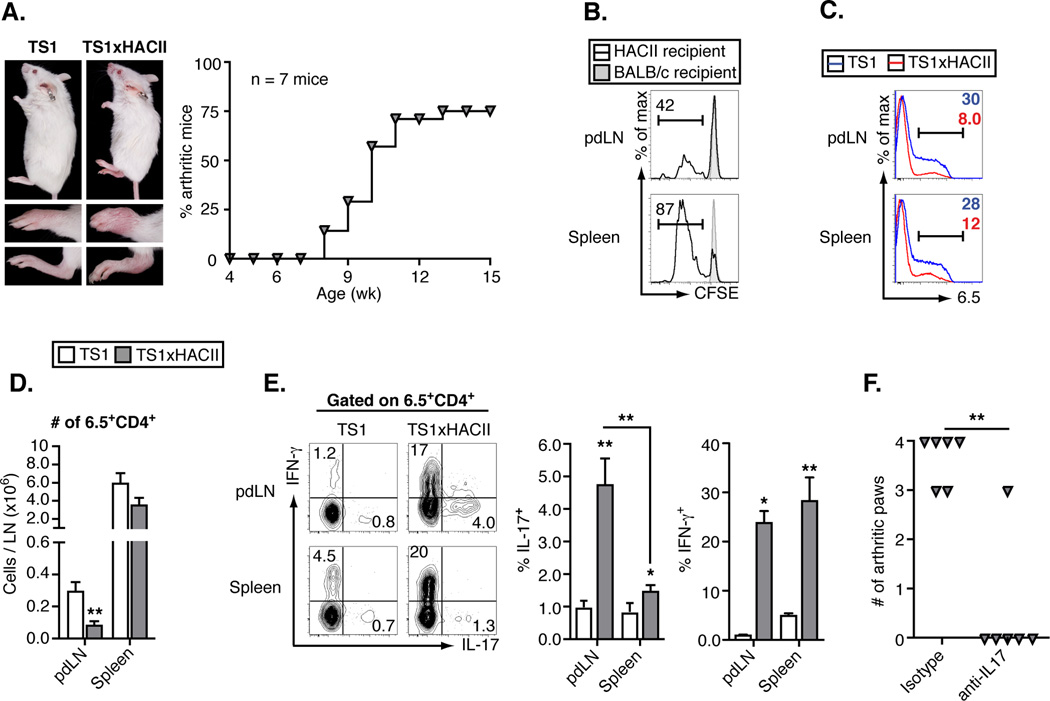
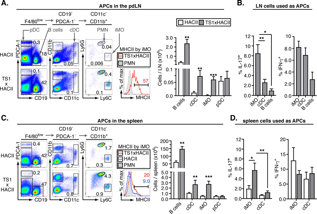
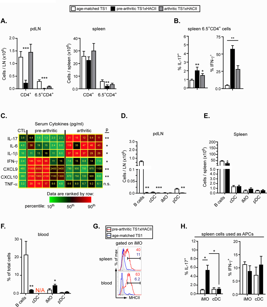
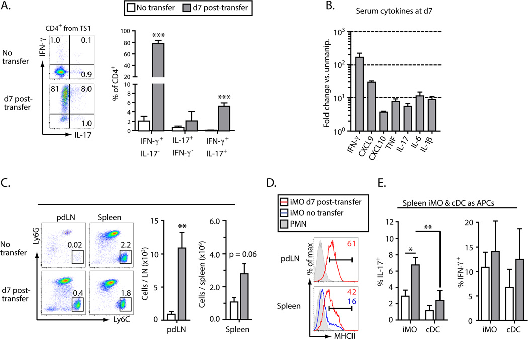
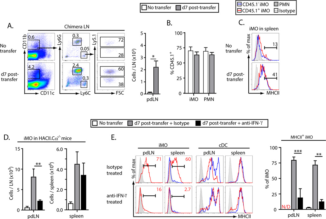
We have examined mechanisms underlying the formation of pathologic Th17 cells using a transgenic mouse model in which autoreactive CD4(+) T cells recognize influenza virus hemagglutinin (HA) as a ubiquitously expressed self-Ag and induce inflammatory arthritis. The lymph nodes of arthritic mice contain elevated numbers of inflammatory monocytes (iMO) with an enhanced capacity to promote CD4(+) Th17 cell differentiation, and a regional inflammatory response develops in the paw-draining lymph nodes by an IL-17-dependent mechanism. The activation of these Th17-trophic iMO precedes arthritis development and occurs in the context of an autoreactive CD4(+) Th1 cell response. Adoptive transfer of HA-specific CD4(+) T cells into nonarthritic mice expressing HA as a self-Ag similarly led to the formation of Th1 cells and of iMO that could support Th17 cell formation, and, notably, the accumulation of these iMO in the lymph nodes was blocked by IFN-γ neutralization. These studies show that autoreactive CD4(+) Th1 cells directed to a systemically distributed self-Ag can promote the development of a regional Th17 cell inflammatory response by driving the recruitment of Th17-trophic iMO to the lymph nodes.


Figures

Figure 1
Figure 1
Arthritis in TS1xHACII mice is accompanied by a regional Th17 response to a systemic self-antigen. (A) Photographs of age and sex-matched TS1 and arthritic TS1xHACII mice, lower panels show front and rear paws. Graph shows arthritis incidence in a cohort of TS1xHACII mice. (B) CFSE dilution by CD4+ T cells from TS1 mice 3 d post-transfer into HACII or BALB/c recipients. (C) Histograms showing 6.5 staining by CD4+ T cells in pdLN and spleens of TS1 and arthritic TS1xHACII mice. (D) Numbers of 6.5+CD4+ T cells in pdLN and spleens of TS1 and arthritic TS1xHACII mice. (E) Ex vivo production of IFN-γ and IL-17 by 6.5+CD4+ T cells from pdLN and spleens of TS1 and arthritic TS1xHACII mice. (F) Arthritis incidence in 15 week old TS1xHACII mice given weekly injections of anti-IL-17 beginning at 4 wk of age. All data are means ± SEM of ≥ 5 independent determinations. *p<0.05; **p<0.01.
Figure 2
Figure 2
Th17-trophic inflammatory monocytes accumulate in arthritic TS1xHACII mice. (A) Representative flow data showing APC subsets in the pdLN. The percent of total live cells falling within each gate is indicated. MHCII expression by iMO and the total numbers of each APC are shown in the accompanying panels. (B) Percentage of 6.5+CD4+ T cells that produced IL-17 or IFN-γ following 7 days of co-culture with APC subsets purified from the LN of arthritic TS1xHACII mice. (C) As in A, except for spleen. (D) As in B, except co-culture was with APC subsets purified from the spleens of HACII mice or arthritic TS1xHACII mice. All data are means ± SEM of ≥ 5 independent determinations. *p<0.05; **p<0.01; ***p<0.005.
Figure 3
Figure 3
A systemic CD4+ Th1 cytokine response precedes arthritis in TS1xHACII mice. (A) Number of CD4+ and 6.5+CD4+ T cells in the pdLN and spleens of TS1, pre-arthritic TS1xHACII, and arthritic TS1xHACII mice. (B) Percentage of 6.5+CD4+ splenocytes that produced IL-17 or IFN-γ (C) Heat map showing the concentrations of Th17 and Th1 cytokines in the serum of pre-arthritic and arthritic TS1xHACII mice. Average values from 7 control mice are shown in the leftmost column. The concentration of each cytokine is indicated within the tiles. P values refer to comparisons between pre-arthritic and arthritic TS1xHACII mice. (D–F) Number of APCs in the pdLN (D) and spleen (E), and percentages of cells in the blood (F) of 5 wk-old TS1xHACII and TS1 mice. N/A = equivalent subset not present. (G) MHCII expression by iMO in the spleens and blood of 5 wk-old TS1xHACII and TS1 mice. (H) Percentage of 6.5+CD4+ T cells from TS1 mice that produced IL-17 or IFN-γ following 7 d of co-culture with APC subsets purified from the spleens of 5 wk old TS1xHACII or HACII mice. All data are means ± SEM of N ≥ 5 independent determinations. *p<0.05; **p<0.01; ***p<0.005.
Figure 4
Figure 4
Autoreactive T cells promote the activation of Th17-trophic inflammatory monocytes. CD4+ T cells from TS1 mice were transferred into HACII.Cα−/− mice (“recipient mice”). All data were collected at 7 d post-transfer. (A) Production of IFN-γ and IL-17 by CD4+ T cells from a TS1 mouse analyzed directly ex vivo or at 7 d post-transfer. (B) Fold increase in the indicated cytokines in the serum of recipient versus unmanipulated mice. (C) Representative flow data of iMO and PMN in the pdLN and spleens. The frequency of each subset as a percentage of total cells is indicated on each plot. The total number of iMO in the spleens and pdLN of recipient mice is shown in the accompanying chart. (D) Histograms showing MHCII expression by iMO in the spleens and pdLN of recipient mice. (E) Percentages of 6.5+CD4+ T cells that produced IL-17 or IFN-γ following 7 d of co-culture with APCs isolated from the spleens of unmanipulated HACII or recipient mice. All data are means ± SEM of N ≥ 3 independent determinations. *p<0.05; **p<0.01; ***p<0.005.
Figure 5
Figure 5
IFN-γ promotes in vivo activation of Th17-trophic inflammatory monocytes. (A–C) Lethally irradiated BALB/c mice were reconstituted with mixed bone marrow from HACII.CD45.1+ (75%) and BALB/c.CD45.1 (25%) mice. Four weeks after reconstitution CD4+ T cells from a TS1 mouse were adoptively transferred into the chimeras and data were collected at 7 d post-transfer. (A) Representative flow data showing the accumulation of iMO in the pdLN of chimeric mice at d7 post-transfer. Gating for live CD19PDCA-1 cells is shown. (B) The frequency of iMO and PMN that were CD45.1+ in the pdLN of chimeric mice at d7 post-transfer. (C) Representative histograms showing MHCII expression by iMO and PMN in the spleens of chimeric mice at d7 post-transfer. Flow data are representative of 3 independent experiments with at least one mouse per condition per experiment. (F–G) As in Figure 4 except mice were treated at d0 and d3 with an anti-IFN-γ neutralizing mAb. (F) Number of iMO in the pdLN of recipient mice treated with anti-IFN-γ or an isotype control mAb. (G) Histograms showing MHCII expression by iMO and cDC in the spleens and pdLN of recipient mice treated with anti-IFN-γ or an isotype control mAb. All numerical data are means ± SEM of at least 3 independent experiments.

References

    1. Korn T, Bettelli E, Oukka M, Kuchroo VK. IL-17 and Th17 Cells. Annu. Rev. Immunol. 2009;27:485–517. - PubMed
    1. Peck A, Mellins ED. Precarious balance: Th17 cells in host defense. Infect. Immun. 2010;78:32–38. - PMC - PubMed
    1. Mangan PR, Harrington LE, O’Quinn DB, Helms WS, Bullard DC, Elson CO, Hatton RD, Wahl SM, Schoeb TR, Weaver CT. Transforming growth factor-beta induces development of the T(H)17 lineage. Nature. 2006;441:231–234. - PubMed
    1. Bettelli E, Carrier Y, Gao W, Korn T, Strom TB, Oukka M, Weiner HL, Kuchroo VK. Reciprocal developmental pathways for the generation of pathogenic effector TH17 and regulatory T cells. Nature. 2006;441:235–238. - PubMed
    1. McGeachy MJ, Chen Y, Tato CM, Laurence A, Joyce-Shaikh B, Blumenschein WM, McClanahan TK, O’Shea JJ, Cua DJ. The interleukin 23 receptor is essential for the terminal differentiation of interleukin 17-producing effector T helper cells in vivo. Nat. Immunol. 2009;10:314–324. - PMC - PubMed

Publication types