

The importance of microglia in the development of the vasculature in the central nervous system
- PMID: 23422217
- PMCID: PMC3583711
- DOI: 10.1186/2045-824X-5-4
The importance of microglia in the development of the vasculature in the central nervous system
Erratum in
-
Correction: The importance of microglia in the development of the vasculature in the central nervous system.Vasc Cell. 2013 Jun 25;5(1):12. doi: 10.1186/2045-824X-5-12. Vasc Cell. 2013. PMID: 23809768 Free PMC article.
Abstract
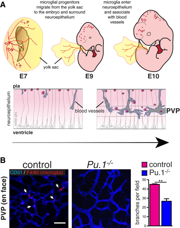
The body's vascular system is thought to have developed in order to supply oxygen and nutrients to cells beyond the reach of simple diffusion. Hence, relative hypoxia in the growing central nervous system (CNS) is a major driving force for the ingression and refinement of the complex vascular bed that serves it. However, even before the establishment of this CNS vascular system, CNS-specific macrophages (microglia) migrate into the brain. Recent studies in mice point to the fundamental importance of microglia in shaping CNS vasculature during development, and re-shaping these vessels during pathological insults. In this review, we discuss the origin of CNS microglia and their localization within the brain based on data obtained in mice. We then review evidence supporting a functional role of these microglia in developmental angiogenesis. Although pathologic processes such as CNS ischemia may subvert the developmental functions of microglia/macrophages with significant effects on brain neo-angiogenesis, we have left this topic to other recent reviews (Nat Rev Immunol 9:259-270, 2009 and Trends Mol Med 17:743-752, 2011).
Figures
References
LinkOut - more resources
Full Text Sources
Other Literature Sources
