. 2013:4:1494.
doi: 10.1038/ncomms2493.

Parasympathetic stimulation improves epithelial organ regeneration

Affiliations

Parasympathetic stimulation improves epithelial organ regeneration

Sarah M Knox et al. Nat Commun. 2013.

Abstract

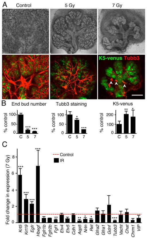
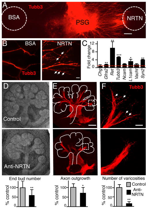
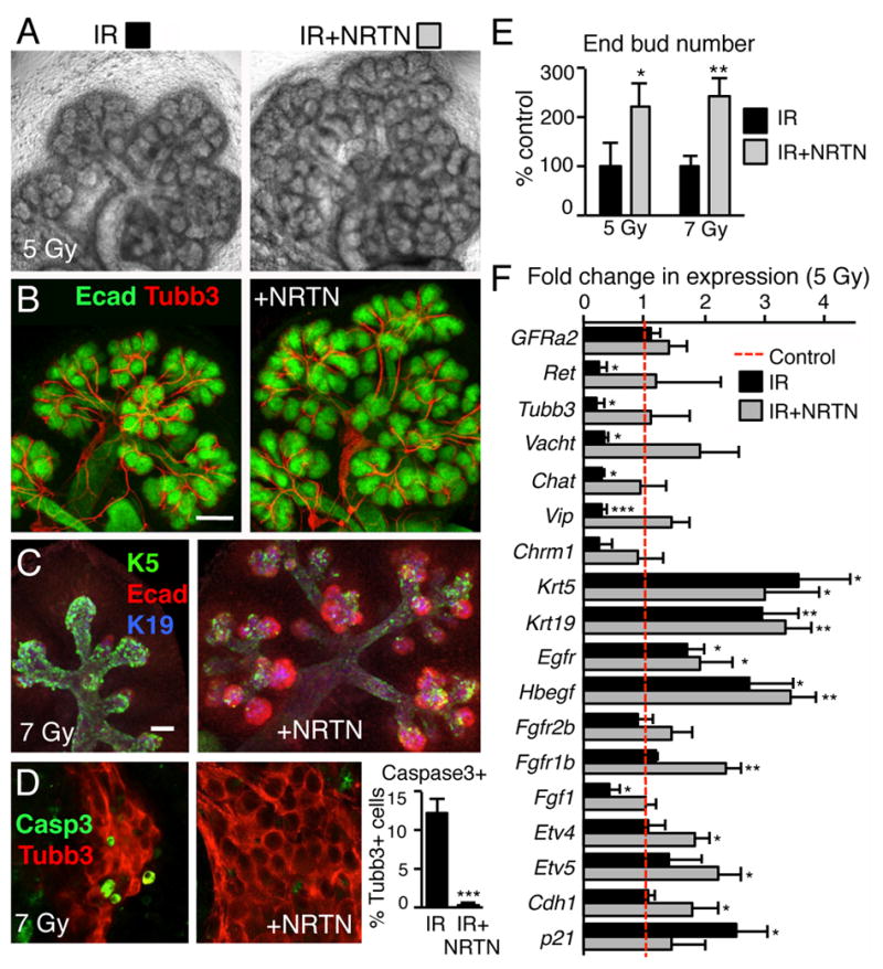
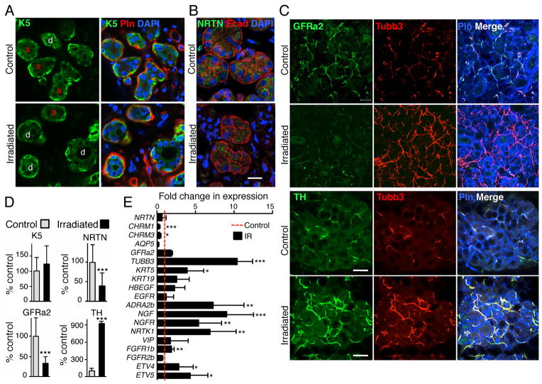
Parasympathetic nerves are a vital component of the progenitor cell niche during development, maintaining a pool of progenitors for organogenesis. Injured adult organs do not regenerate after parasympathectomy, and there are few treatments to improve organ regeneration, particularly after damage by therapeutic irradiation. Here we show that restoring parasympathetic function with the neurotrophic factor neurturin increases epithelial organ regeneration after damage. We use mouse salivary gland explant culture containing fluorescently labelled progenitors, and injure the tissue with irradiation. The progenitors survive, parasympathetic function is diminished and epithelial apoptosis reduces the expression of neurturin, which increases neuronal apoptosis. Treatment with neurturin reduces neuronal apoptosis, restores parasympathetic function and increases epithelial regeneration. Furthermore, adult human salivary glands damaged by irradiation also have reduced parasympathetic innervation. We propose that neurturin will protect the parasympathetic nerves from damage and improve organ regeneration. This concept may be applicable for other organs where parasympathetic innervation influences their function.


Conflict of interest statement

Competing financial interests: The authors declare no competing financial interests.

Figures

Figure 1
Figure 1. Irradiation decreases epithelial morphogenesis and parasympathetic innervation
(A) Submandibular glands (SMG) from E13 K5-venus transgenic mice irradiated (IR dose of 5 and 7 Gy) and cultured for 5 days. Whole-mount images of K5-venus and nerves (Tubb3) are shown. Arrows point to ducts and arrowheads point to small ganglia. Scale bar, 200 μM. (B) Quantitation of epithelial morphogenesis (End bud number), innervation (Tubb3 fluorescence), and K5+ progenitors (K5-venus fluorescence) were quantitated and normalized to the control (C = 100%). 3–5 SMGs were used/group and experiments repeated 3 times, see SOM for details. ANOVAwith post hoc Dunnett’s test; ***P < 0.001, **P < 0.01, *P < 0.05. (C) qPCR analysis of gene expression in E13 SMGs 3 days after 7 Gy IR compared to control. Means ± SD from three experiments. A False Discovery Rate (Q) for multiple unpaired t tests was set to 5%; ***P < 0.001, **P < 0.01, *P < 0.025.
Figure 2
Figure 2. NRTN increases parasympathetic neurite outgrowth and function
(A) Whole mount image (Tubb3 staining) of a parasympathetic ganglia (PSG) cultured with a BSA and a NRTN-coated bead. Scale bar, 100 μm. (B) Higher magnification of neurites to show varicosities (arrows). Scale bar, 20 μm. (C) QPCR analysis of gene expression in PSGs treated with NRTN. Data was normalized to Rsp29 and BSA-treated PSGs. (D) Images of SMGs cultured with a function-blocking NRTN antibody or an IgG control. Epithelial morphogenesis (end bud number) was quantitated and normalized to IgG control. (E) Whole mount staining of the nerves. Axon outgrowth was measured using Tubb3 fluorescence and normalized to epithelial area, see SOM for details. Scale bar, 100 μm. (F) Higher magnification of nerves highlights the varicosities. The number of varicosities was counted. Scale bar, 20 μm. All graphs are all normalized to the IgG control. Means ± SD from three experiments. Student’s t test; ***P < 0.001, **P < 0.01, *P < 0.05.
Figure 3
Figure 3. NRTN treatment reduces neuronal apoptosis and restores parasympathetic function
(A) SMGs from E14 mice were irradiated with 5 Gy, treated with NRTN (1 ng/ml), and cultured for 3 days. (B) Whole-mount images of the epithelium (Ecad) and nerves (Tubb3) are shown. Scale bar, 100 μm. (C) E14 SMGs were irradiated with 7 Gy, cultured 3 days and stained for epithelium (Ecad), K5+ cells and K19+ cells (also see Supplementary Fig. S5A). Scale bar, 50 μm. (D) Neuronal apoptosis was measured in E14 SMGs treated with 7 Gy IR for 3 days by caspase3 staining and the percentage of Casp3+Tubb3+ cells measured. Means ± SD from three experiments. Student’s t test; ***P < 0.001. (E) Epithelial morphogenesis (end bud number) was quantitated and normalized to IR SMG. Means ± SD from three experiments. Student’s t test; **P < 0.01, *P < 0.05. (F) qPCR analysis after 3 days culture of Control and 5 Gy IR +/− NRTN treatment. Data was normalized to Rsp29 and non-IR control. Mean ± SD of three independent experiments. ANOVA with post hoc Dunnett’s test; *P < 0.05, **P < 0.01, ***P < 0.001.
Figure 4
Figure 4. IR of adult human SMGs reduces parasympathetic innervation
(A) Control non-IR and IR SMGs were immunostained for K5; the basement membrane was stained with perlecan (Pln), and the nuclei with DAPI. Acinar units (a) and ducts (d) are labeled in the K5 images. (B) NRTN immunostaining is in the epithelium (Ecad). Nuclei are stained with DAPI. Images are single confocal sections (2 μm) Scale bar, 20 μm. (C) GFRa2 immunostaining labels the parasympathetic nerves, tyrosine hydroxylase (TH) labels sympathetic nerves Perlecan (Pln) labels the basement membrane, and Tubb3 is a pan-neuronal marker. Images are single confocal projections (20 μm), scale bar, 50 μm. (D) K5, NRTN, GFRa2 and TH fluorescence was quantitated, K5, GFRa2 and TH were normalized to perlecan, which stains IR and control tissues at a similar level, but NRTN was normalized to Ecad to show that the remaining epithelium produces less NRTN, and all were expressed as a percentage of non-IR control. (E) qPCR analysis of gene expression normalized to GAPDH and non-IR control. Mean ± SD of IR (n=3) and non-IR human SMGs (n=5). A False Discovery Rate (Q) for multiple unpaired t tests was set to 5%; ***P < 0.001, **P < 0.01, *P < 0.025.

References

    1. Bergmann A, Steller H. Apoptosis, stem cells, and tissue regeneration. Sci Signal. 2010;3(145):re8. - PMC - PubMed
    1. King RS, Newmark PA. Beyond the cell: The cell biology of regeneration. J Cell Biol. 2012;196(5):553–562. - PMC - PubMed
    1. Carlson BM. Some principles of regeneration in mammalian systems. Anat Rec B New Anat. 2005;287(1):4–13. - PubMed
    1. Proctor GB, Carpenter GH. Regulation of salivary gland function by autonomic nerves. Auton Neurosci. 2007;133(1):3–18. - PubMed
    1. Tatsuishi Y, Hirota M, Kishi T, Adachi M, Fukui T, Mitsudo K, et al. Human salivary gland stem/progenitor cells remain dormant even after irradiation. Int J Mol Med. 2009;24(3):361–366. - PubMed

Publication types

MeSH terms