. 2013 Mar 28;54(3):1898-904.
doi: 10.1167/iovs.12-11375.

A new mouse model of inducible, chronic retinal ganglion cell dysfunction not associated with cell death

Affiliations

A new mouse model of inducible, chronic retinal ganglion cell dysfunction not associated with cell death

Xu Yang et al. Invest Ophthalmol Vis Sci. .

Abstract

Purpose: To develop a mouse model of inducible, chronic retinal ganglion cell (RGC) dysfunction not associated with cell death.

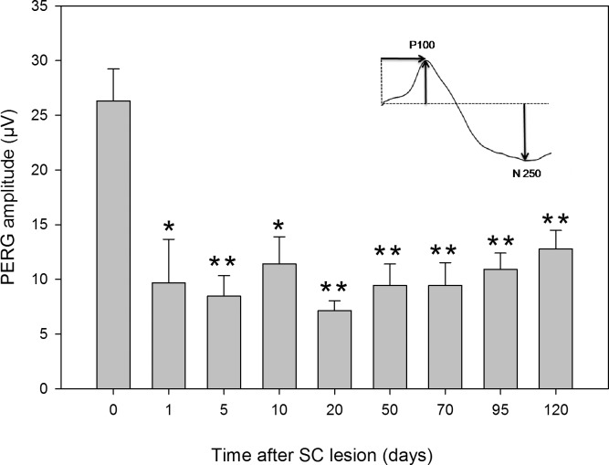
Methods: Eighteen C57BL/6J mice were longitudinally tested with pattern electroretinogram (PERG) and spectral-domain optical coherence tomography (OCT) before and after aspiration of the contralateral superior colliculus (SC), which removed terminals of optic tract axons and the superficial layers of the SC. At the 4-month end points, retinas were harvested for Brn3b immunostaining and BDNF immunoblotting.

Results: The PERG lost approximately 60% of its baseline amplitude (P < 0.01) within the first day after lesion, and remained at a reduced level over 4 months. At the end point, the density of Brn3b-positive RGCs was normal, but their nucleus size was reduced by approximately 24% (P < 001). OCT measurements showed thinning of the inner, but not outer, retina by approximately 9% (P < 0.01) starting 10 to 20 days after lesion. Retinal nerve fiber layer thickness was unchanged. At the end point, retinal homogenates showed a substantial overexpression of BDNF protein level.

Conclusions: Mechanical SC lesion in adult mice results in a rapid, chronic loss of RGC electrical responsiveness that is followed by cell shrinkage but not cell death. The SC-lesion mouse represents a new, inducible model that allows investigating stages and mechanisms of RGC dysfunction without the confounding effects of cell death that are common in the existing models of optic neuropathies and optic nerve lesions.


Conflict of interest statement

Disclosure: X. Yang, None; T.-H. Chou, None; M. Ruggeri, None; V. Porciatti, None

Figures

Figure 1
Figure 1
PERG amplitude before and at different times after lesion of the contralateral SC. Lesion was done on the right SC, and the PERG was recorded from the left eye. Error bars represent the SEM (n = 18). The inset shows a representative example of PERG response, whose amplitude was measured from the peak of the P100 wave to the trough of the N250 wave. Asterisks above the bars represent the level of significance compared to baseline (*P < 0.05; **P < 0.01).
Figure 2.
Figure 2.
Mean thickness of different retinal layers before and after lesion of the contralateral SC. (A) Inner retina, (B) outer retina, (C) retinal nerve fiber layer (RNFL). Error bars represent the SEM (n = 8). Asterisks above the gray bars represent the level of significance compared with baseline (**P < 0.01). (D) Example of one OCT image intersecting the optic nerve head at its center. Vertical bars represent retinal layers that have been measured (short bar: inner retina; longer bar: total retina; horizontal parallel bars: retinal fiber layer). (E, F) Segmentation of inner retina thickness (E) compared with total retina thickness (F) for a hollow cylinder of tissue (outer radius 0.65 mm, inner radius 0.2 mm) centered on the optic disk. Images were obtained from a raster of 100 b-scans spanning an area of 1.3 × 1.3 mm centered on the optic disk. RNFL thickness was manually measured on a horizontal slice of tissue crossing the center of the optic disk and at an eccentricity of 275 μm for the disk center.
Figure 3.
Figure 3.
Cell density (A) and nucleus size (B) of Brn3b-positive RGCs obtained in control retinas and in retinas of eyes that had their contralateral SC lesioned 4 months earlier. Error bars represent the SEM (n = 6). The asterisk above the bars represents the level of significance compared with baseline (*P < 0.05). (C, D) representative examples of Brn3b-positive RGCs in control (C) and SC-lesioned retinas (D). The calibration bar in (C) represents 50 μm.
Figure 4.
Figure 4.
Increase in retinal BDNF expression in response to aspiration of the superficial layers of the contralateral SC 4 months earlier. Western blot analysis was conducted on individual retinal homogenates (n = 5) and compared with one homogenate of pooled control retinas (n = 6). (A) Western blots of BDNF compared with β-actin control. (B) Normalized BDNF/β-actin ratios obtained by densitometric analysis in pooled control retinas and in retinas of SC-lesioned mice. Double asterisks above the right bar indicate that in SC-lesioned retinas, BDNF was significantly overexpressed (P < 0.01). Error bars represent the ±95% confidence limits of the mean.

References

    1. Levkovitch-Verbin H. Animal models of optic nerve diseases. Eye. 2004; 18: 1066–1074 - PubMed
    1. Pang IH, Clark AF. Rodent models for glaucoma retinopathy and optic neuropathy. J Glaucoma. 2007; 16: 483–505 - PubMed
    1. Buckingham BP, Inman DM, Lambert W, et al. Progressive ganglion cell degeneration precedes neuronal loss in a mouse model of glaucoma. J Neurosci. 2008; 28: 2735–2744 - PMC - PubMed
    1. Howell GR, Libby RT, Jakobs TC, et al. Axons of retinal ganglion cells are insulted in the optic nerve early in DBA/2J glaucoma. J Cell Biol. 2007; 179: 1523–1537 - PMC - PubMed
    1. Saleh M, Nagaraju M, Porciatti V. Longitudinal evaluation of retinal ganglion cell function and IOP in the DBA/2J mouse model of glaucoma. Invest Ophthalmol Vis Sci. 2007; 48: 4564–4572 - PMC - PubMed

Publication types