. 2013;11(2):e1001479.
doi: 10.1371/journal.pbio.1001479. Epub 2013 Feb 12.

HAMP domain conformers that propagate opposite signals in bacterial chemoreceptors

Affiliations

HAMP domain conformers that propagate opposite signals in bacterial chemoreceptors

Michael V Airola et al. PLoS Biol. 2013.

Abstract

HAMP domains are signal relay modules in >26,000 receptors of bacteria, eukaryotes, and archaea that mediate processes involved in chemotaxis, pathogenesis, and biofilm formation. We identify two HAMP conformations distinguished by a four- to two-helix packing transition at the C-termini that send opposing signals in bacterial chemoreceptors. Crystal structures of signal-locked mutants establish the observed structure-to-function relationships. Pulsed dipolar electron spin resonance spectroscopy of spin-labeled soluble receptors active in cells verify that the crystallographically defined HAMP conformers are maintained in the receptors and influence the structure and activity of downstream domains accordingly. Mutation of HR2, a key residue for setting the HAMP conformation and generating an inhibitory signal, shifts HAMP structure and receptor output to an activating state. Another HR2 variant displays an inverted response with respect to ligand and demonstrates the fine energetic balance between "on" and "off" conformers. A DExG motif found in membrane proximal HAMP domains is shown to be critical for responses to extracellular ligand. Our findings directly correlate in vivo signaling with HAMP structure, stability, and dynamics to establish a comprehensive model for HAMP-mediated signal relay that consolidates existing views on how conformational signals propagate in receptors. Moreover, we have developed a rational means to manipulate HAMP structure and function that may prove useful in the engineering of bacterial taxis responses.


Conflict of interest statement

The authors have declared that no competing interests exist.

Figures

Figure 1
Figure 1. Schematic of Aer2-Tar Chimeras.
The HAMP domain of Tar was replaced with single and poly-HAMP domains from Aer2 to generate chimeric receptors. Transmembrane ATCs (e.g., H1) contained the ligand binding domain and Tar KCM, both of which are necessary for modulating CheA kinase activity in response to aspartate. Soluble ATCs (e.g., H1s) comprised fusions of the Aer2 HAMP domains with only the Tar KCM. The structure of the three-unit Aer2 poly-HAMP domain (Protein Data Bank code 3LNR) is shown on the right, with HAMP1 (blue), HAMP2 (yellow/orange), and HAMP3 (purple) colored accordingly.
Figure 2
Figure 2. Signaling biases and expression levels of ATC receptors.
(A) Structures of HAMP1 and HAMP2, highlighting positions of mutations reported in this study. HR2 (I88G) plays a prominent role in the HAMP2 hydrophobic core, inserting into the HAMP bundle between AS1 and AS2, while HR2 (V33G) in HAMP1 appears dispensable for bundle stability as it resides on the domain periphery. L21 and L44 occupy core heptad positions inside the HAMP bundle. Membrane-associated HAMP domains contain a highly conserved DExG motif at the connector-AS2 junction and a less conserved Pro residue between TM2 and AS1. (B) Tumbling biases of transmembrane and soluble ATC receptors quantified by temporal assays in CheRB+ and CheRB− cells. Signaling biases are grouped into four categories: (1) CCW locked (<5% CW), (2) slight CW bias (5%–10% CW), (3) CW bias (10%–50% CW), and (4) strong CW bias (50%–95% CW) or CW locked (>95% CW). Temporal assays confirm H1 and H1-2 induce opposite outputs. The L44H mutation generates a CW locked receptor with or without the adaptation system. The soluble receptors H1s and H1-2s generate more distinct CW and CCW locked phenotypes in CheRB− cells than their transmembrane counterparts. Mutation of HR2 in H1-2s I88G switches receptor signaling from CCW to CW locked, which is consistent with HR2 stabilizing the CCW HAMP2 conformer. (C) Expression levels of ATC receptors in CheRB+ (BT3388) cells, normalized to that of Tar for transmembrane receptors and that of Tar KCM for soluble receptors.
Figure 3
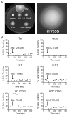
Figure 3. H1D and H1 V33G receptors both respond to attractant, but with normal and inverse responses, respectively.
(A) Swim assays of ATC receptors on tryptone agar plates. Colonies with functional chemoreceptors generate a characteristic ring near the leading edge of an expanding colony as cells consume Asp and swim towards higher Asp concentrations. H1 V33G generates an inverted ring, in comparison to Tar, which suggests an inverted CCW-to-CW response to Asp. (B) Temporal assays of transmembrane receptors showing response and adaptation kinetics. CheRB+ cells expressing various receptors were allowed to reach adaptation equilibrium before Asp was added. Tumbling frequencies alter if receptors are capable of receiving and relaying signal input from TM2 to the output KCM. Tar responds in the normal direction, switching from 12.5% to 1% CW bias. After ∼300 s, the adaptation system restores Tar CW bias to 12.5%. H1D has a normal Asp response, switching from 17.5% to 2.5% CW bias. H1 V33G displays an inverted response, switching from 16% to 100% CW bias upon Asp addition. A lower concentration of Asp is representative of increased receptor sensitivity.
Figure 4
Figure 4. Structure of L44H and V33G mutants.
(A) Crystal structure of Aer2 1–172 proteins, with HAMP1 colored blue (WT), purple (L44H), and green (V33G). (B) Superposition of WT HAMP1 and CW locked L44H HAMP1 mutant. The L44H side chain redirects from the bundle core towards AS1, causing a 15° tilt of AS1 away from AS2 and a 5 Å shift at the top of AS1, which disrupts the upstream helical coiled coil. The positions of the AS2 output helices are identical to WT, which confirms that a HAMP1 structure generates CW output. (C) Superposition of HAMP1 in WT and in the inverted signaling V33G mutant. Removal of HR2 in V33G does not affect the helical position of AS1 or AS2, suggesting that HR2 is dispensable to generate a HAMP1 conformer. 2Fo-Fc electron density maps of V33G (contoured at 2 σ) lack density in the surrounding HR2 connector region, suggesting increased flexibility of this region. Increased flexibility of HR2 due to Gly substitution would destabilize a HAMP2 conformer and thereby favor CW output.
Figure 5
Figure 5. Conformational properties of soluble receptors assessed by PDS.
(A) Schematic of spin-label sites in Aer2 1–172 (H1C and H2C) and soluble ATCs (H1s and H1-2s). Sites were chosen in AS1 and AS2 to maximize the distance separation expected to distinguish HAMP1 and HAMP2 in the crystal structure. Cα–Cα distances from the crystal structure are shown (top distance). MTSSL spin labels can add up to 13 Å (bottom distance). (B) Inter-spin distances measured by PDS of spin-labeled proteins. Pair-wise distance distributions (P[r]) of control samples (H1C and H2C) matched well with the differences in the crystal structure (Table 1). Attachment to the Tar KCM (H1s) results in a more dynamic HAMP1 conformer, with broad distance distributions, which is indicative of conformation exchange between HAMP1 and HAMP2. HAMP2, in H1-2s, remains relatively static, with narrow distance peaks that are nearly identical to those of H2C. The H1-2s I88G HR2 mutant switches the conformational properties of HAMP2 towards a dynamic HAMP1 state, consistent with the CW locked phenotype in vivo. The two HAMP conformers have opposite effects downstream. HAMP2, which forms a two-helix coiled coil at the end of AS2, maintains similar distances across the junction in the KCM. HAMP1, which forms a four-helix coiled coil, maintains a longer, broader distance distribution. This suggests the structure of the AS2 helices is propagated downstream into the KCM helical bundle. Inter-spin distance distributions are tabulated in Table 1.
Figure 6
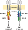
Figure 6. Model for HAMP domain signal relay in bacterial chemoreceptors.
The HAMP domains of MCPs exchange between HAMP1 and HAMP2 states to regulate bacterial chemotaxis. The conformation of HAMP2 imparts a two-helix coiled coil across the AS2/KCM junction, which results in CheA kinase inhibition and CCW flagella rotation. A dynamic HAMP1 forms a continuous four-helix coiled coil across the junction to generate kinase activation and CW flagella rotation.

Comment in

References

    1. Szurmant H, White RA, Hoch JA (2007) Sensor complexes regulating two-component signal transduction. Curr Opin Struct Biol 17: 706–715. - PMC - PubMed
    1. Letunic I, Doerks T, Bork P (2012) SMART 7: recent updates to the protein domain annotation resource. Nucleic Acids Res 40: D302–D305. - PMC - PubMed
    1. Parkinson JS (2010) Signaling mechanisms of HAMP domains in chemoreceptors and sensor kinases. Annu Rev Microbiol 64: 101–122. - PubMed
    1. Hazelbauer GL, Falke JJ, Parkinson JS (2008) Bacterial chemoreceptors: high-performance signaling in networked arrays. Trends Biochem Sci 33: 9–19. - PMC - PubMed
    1. Hulko M, Berndt F, Gruber M, Linder JU, Truffault V, et al. (2006) The HAMP domain structure implies helix rotation in transmembrane signaling. Cell 126: 929–940. - PubMed

Publication types

MeSH terms

Substances

LinkOut - more resources