. 2013;11(2):e1001482.
doi: 10.1371/journal.pbio.1001482. Epub 2013 Feb 12.

Reciprocal regulation of protein synthesis and carbon metabolism for thylakoid membrane biogenesis

Affiliations

Reciprocal regulation of protein synthesis and carbon metabolism for thylakoid membrane biogenesis

Alexandra-Viola Bohne et al. PLoS Biol. 2013.

Abstract

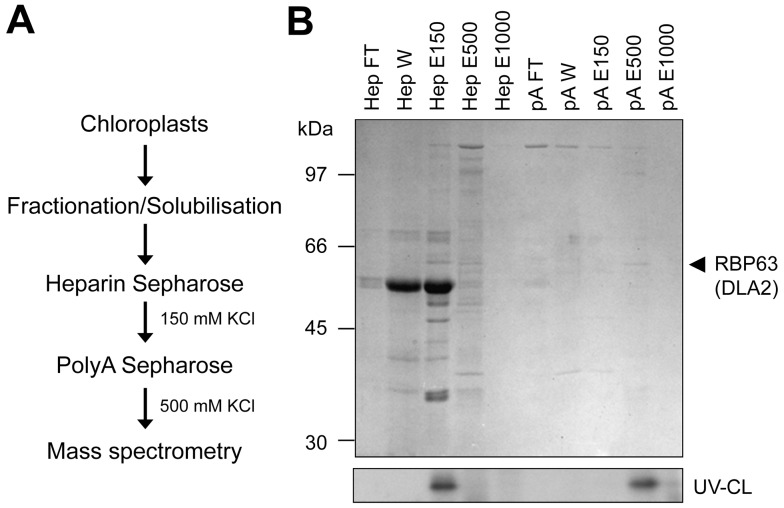
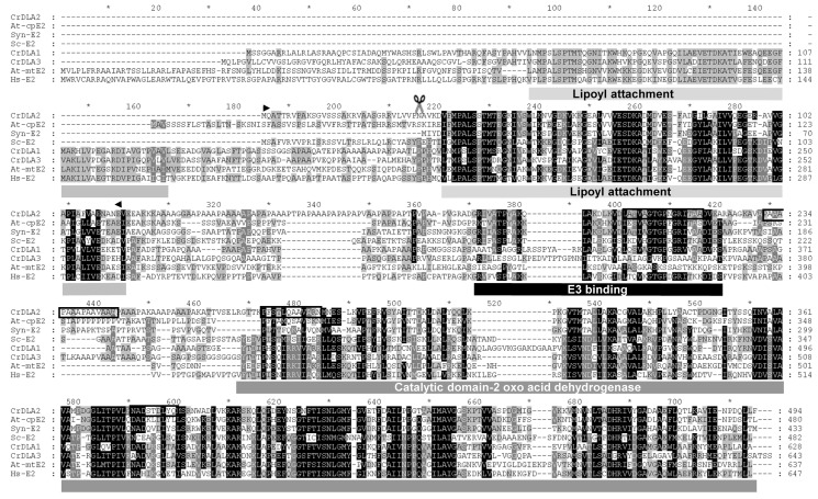
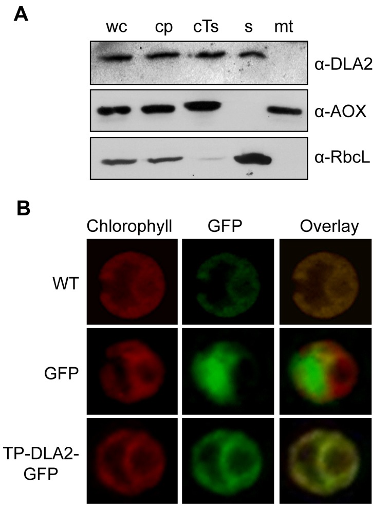
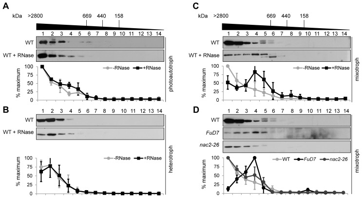
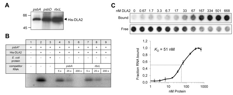
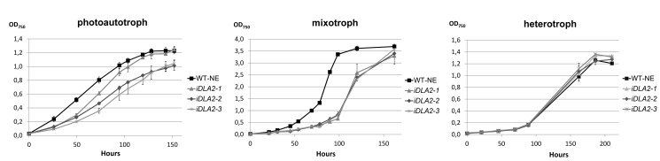
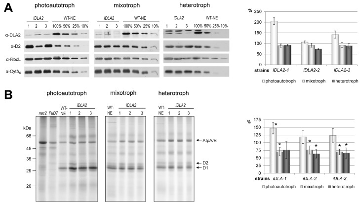
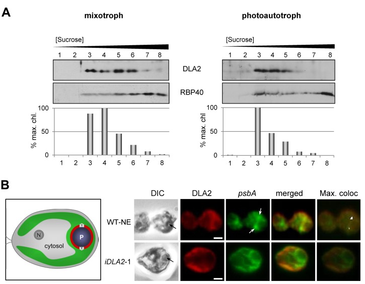
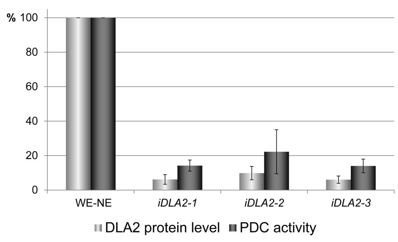
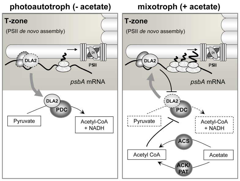
Metabolic control of gene expression coordinates the levels of specific gene products to meet cellular demand for their activities. This control can be exerted by metabolites acting as regulatory signals and/or a class of metabolic enzymes with dual functions as regulators of gene expression. However, little is known about how metabolic signals affect the balance between enzymatic and regulatory roles of these dual functional proteins. We previously described the RNA binding activity of a 63 kDa chloroplast protein from Chlamydomonas reinhardtii, which has been implicated in expression of the psbA mRNA, encoding the D1 protein of photosystem II. Here, we identify this factor as dihydrolipoamide acetyltransferase (DLA2), a subunit of the chloroplast pyruvate dehydrogenase complex (cpPDC), which is known to provide acetyl-CoA for fatty acid synthesis. Analyses of RNAi lines revealed that DLA2 is involved in the synthesis of both D1 and acetyl-CoA. Gel filtration analyses demonstrated an RNP complex containing DLA2 and the chloroplast psbA mRNA specifically in cells metabolizing acetate. An intrinsic RNA binding activity of DLA2 was confirmed by in vitro RNA binding assays. Results of fluorescence microscopy and subcellular fractionation experiments support a role of DLA2 in acetate-dependent localization of the psbA mRNA to a translation zone within the chloroplast. Reciprocally, the activity of the cpPDC was specifically affected by binding of psbA mRNA. Beyond that, in silico analysis and in vitro RNA binding studies using recombinant proteins support the possibility that RNA binding is an ancient feature of dihydrolipoamide acetyltransferases. Our results suggest a regulatory function of DLA2 in response to growth on reduced carbon energy sources. This raises the intriguing possibility that this regulation functions to coordinate the synthesis of lipids and proteins for the biogenesis of photosynthetic membranes.


Conflict of interest statement

The authors have declared that no competing interests exist.

Figures

Figure 1
Figure 1. Isolation of RBP63.
(A) Flow chart demonstrating the steps used to purify RBP63 (DLA2). (B) SDS-PAGE and Coomassie Blue staining of proteins at various stages of purification (upper panel) and UV cross-linking (UV-CL) of RBP63 to radiolabeled psbA 5′ UTR RNA (bottom panel). HepFT/HepW, flow through/wash fractions from heparin Sepharose; HepE 150, 500, and 1000, eluates obtained with 150, 500, and 1,000 mM KCl from heparin Sepharose, respectively; pA FT/W, flow-through/wash fraction from poly(A)-Sepharose column; pA E150, 550, and 1000, eluates obtained with 150, 500, and 1,000 mM KCl, respectively. The RBP63 protein that eluted with 500 mM KCl from poly(A)-Sepharose is marked by an arrow.
Figure 2
Figure 2. Three homologous dihydrolipoamide-acetyltransferases (DLAs) in C. reinhardtii.
Multiple alignment of deduced amino acid sequences of the three DLA genes of C. reinhardtii along with Synechocystis sp. PCC 6803, human, S. cerevisiae, and A. thaliana sequences. Conserved residues are shaded in black, similar residues in grey. The sequences were aligned with ClustalW, manually edited and visualized using GeneDoc ,. The predicted chloroplast transit peptide of CrDLA2 has a length of 30 amino acids and is indicated by scissors. Functional domains are depicted below the sequences. Boxed regions in the DLA2 amino acid sequence correspond to peptides identified by mass spectrometry. Arrowheads indicate the amino acids fused to GFP (Figure 3B). Accession numbers are as follows: C. reinhardtii CrDLA1 (EDP01882), CrDLA3 (XP_001696403), CrDLA2 (model: au5.g10333 annotated in JGI v4; Joint Genome Institute; http://genome.jgi-psf.org/Chlre4/Chlre4.home.html); A. thaliana At-mtE2 (LTA3, AAV97810), At-cpE2 (LTA2, NP_189215); Synechocystis sp. 6803 Syn-E2 (sll1841, NP_441936); H. sapiens Hs-E2 (DLAT, NM_001931); and S. cerevisiae Sc-E2 (Lat1, NP_014328).
Figure 3
Figure 3. Plastid localization of DLA2.
(A) Cell subfractionation. Cell fractions were prepared as described in Materials and Methods. We separated 30 µg of each protein fraction by SDS-PAGE and subjected them to immunoblot analysis. The blot was probed with antibodies against DLA2 (α-DLA2), the large subunit of Rubisco (α-RbcL), and the mitochondrial AOX (α-AOX, Agrisera). cp, chloroplasts; cTs, crude thylakoids; mt, mitochondria; s, stroma; wc, whole cells. (B) Analysis of the accumulation of DLA2–GFP fusion proteins in the transformed algal UVM4 strain by laser-scanning microscopy. Chlorophyll autofluorescence (Chlorophyll) and expression of GFP without (GFP) or with an N-terminal fusion of the first 114 aa of DLA2 (TP–DLA2–GFP). The untransformed UVM recipient strain served as control (WT). A merged image of the chlorophyll autofluorescence and GFP signals is shown (Overlay).
Figure 4
Figure 4. DLA2 forms part of a HMW complex that contains the psbA mRNA.
SEC analyses of the DLA2 complex of wild-type CC-406 cells grown under photoautotrophic (A), heterotrophic (B), or mixotrophic (C) conditions along with PSII mutants FuD7 and nac2–26 grown under mixotrophic conditions. (D) Solubilized crude thylakoid proteins (treated with RNase or not) were separated by SEC. Fractions 1 to 14 were subjected to protein gel blot analyses using the DLA2 antiserum. Molecular masses shown at the top were estimated by parallel analysis of high molecular mass calibration markers. Below each panel, a quantitation of DLA2 signal intensities on Western blots for each condition is presented. The quantitation of signals was performed by using AlphaEaseFC software (Alpha Innotech Corp.). For each experiment, the highest amount of DLA2 in the SEC fractions was set to 100%. Mean values and error bars were calculated from at least three independent experiments.
Figure 5
Figure 5. Intrinsic RNA binding activity of recombinant His-DLA2 protein.
(A) UV cross-linking experiment. A total of 200 ng of purified His-DLA2 were analyzed by UV cross-linking using 50–100 kcpm of radiolabeled psbA-, psbD, or rbcL- 5′ UTR RNA probes. (B) Competition experiments. We incubated 10 ng of His-DLA2 protein with radiolabeled psbA-RNA (psbA*) and a 5-, 25-, or 200-fold molar excess of the indicated competitor RNAs representing the 5′ UTRs of the psbA or rbcL mRNA, respectively. To exclude an unspecific RNA binding of contaminating E. coli proteins, we used the same volumes as used for the DLA2 protein of an elution fraction obtained from the bacterial host strain transformed with the empty expression vector served as control (E. coli protein, lane 3). (C) Equilibrium binding constant (K D) of recombinant DLA2 protein. Binding reactions containing 6.7 pM 32P-labeled psbA 5′ UTR RNA and indicated molarities of DLA2 were filtered through stacked nitrocellulose and nylon membranes (upper panel) using a dot blot apparatus according to Ostersetzer et al. . Signal intensities of nitrocellulose-bound protein–RNA complexes (Bound) as well as nylon membrane-bound free RNAs (Free) were quantified. The K D value was determined from three experiments performed with the same DLA2 preparation (lower panel).
Figure 6
Figure 6. Growth curves of iDLA2 lines.
Growth rates were determined for the wild-type strain UVM4 transformed with the empty vector NE537 (WT-NE) and iDLA2 lines (iDLA2-1, -2, -3) under photoautotrophic, mixotrophic, or heterotrophic growth conditions by measuring OD750. Error bars represent 1 standard deviation from the mean based on results from three independent cultures.
Figure 7
Figure 7. D1 accumulation and synthesis in iDLA2 lines under different growth conditions.
(A) D1 protein accumulation. Immunoblot analysis of 40 µg of total cell proteins from indicated iDLA2 lines along with a dilution series of the control strain transformed with the empty vector (WT-NE). Cells were cultured photoautotrophically, mixotrophically, or heterotrophically. Protein gel blot analyses were performed using antibodies raised against the proteins indicated at the left (RbcL, large subunit of Rubisco; Cytb6, cytochrome b6; D2, reaction center protein of PSII). Right panel demonstrates the quantification of immunoblot signals of three independent experiments. Quantification was performed using the AlphaEaseFC software (Alpha Innotech Corp.) by calculating the ratio of D2 over Cytb6. Values obtained for WT-NE were set to 100%. Mean values and standard deviations from three independent experiments are shown. (B) D1 protein synthesis. Membrane proteins of depicted strains were pulse-labeled with 35S sulphate, fractionated in SDS-urea-gels, and analyzed by autoradiography. (Right panel) Quantification of signals was performed by using AlphaEaseFC software (Alpha Innotech Corp.) and calculating the ratio of D1 over AtpA/B. Values obtained for WT-NE were set to 100%. Growth conditions as indicated. Mean values and standard deviations from three (photoautotrophic) or five (mixotrophic/heterotrophic) independent experiments are shown. An asterisk indicates that a mean D1/AtpA/B labeling ratio is significantly different from that of WT-NE (100%) as determined by a one-sample t test (p<0.05).
Figure 8
Figure 8. DLA2-dependent localization of the psbA mRNA to the PSII biogenesis region of the chloroplast (T-zone) and CTMs.
(A) Membranes from cells from either mixotrophic (left panel) or photoautotrophic (right panel) conditions were fractionated by isopycnic sucrose gradient ultracentrifugation, and fractions were analyzed by immunoblot for DLA2 and a marker protein for CTM (RBP40). As a marker for thylakoid membranes, chlorophyll concentration is graphed as the percentage of the maximum value. (B) Cells of either the control strain transformed with the empty vector (WT-NE) or DLA2-deficient (iDLA2-1) were grown under mixotrophic conditions, IF-stained with the α-DLA2-antiserum (red), and FISH-probed for the psbA mRNA (green). Black arrows in differential interference contrast (DIC) images at the left indicate the position of the pyrenoid. White arrows indicate where the psbA FISH signal is localized in T-zones. Pixels with the strongest signals obtained by the program Colocalization Finder (ImageJ) were labeled white (max. confocal). Bars, 1 µm. An illustration of a C. reinhardtii cell shows the nucleus (N), cytosol, and chloroplast (green) with the T-zone (T) and pyrenoid (P).
Figure 9
Figure 9. DLA2 is an active subunit of the chloroplast pyruvate dehydrogenase.
cpPDC activity tests in the same iDLA2 lines grown under photoautotrophic conditions were performed as described in Materials and Methods. The cpPDC activity measured in the wild-type transformed with the empty vector (WT-NE) is designated as 100%. Mean values and standard deviations from three independent experiments are shown. For comparison, DLA2 protein accumulation in iDLA2 lines is shown aside (see also Figure 7A).
Figure 10
Figure 10. Regulation of cpPDC activity.
(A) Regulation of the cpPDC by photosynthetic electron flow. cpPDC activity in cell-wall-deficient wild-type cells (WT) and PSII mutants FuD7 and nac2–26 grown in the presence of acetate under moderate light conditions (L) or in the dark (D). If indicated, wild-type cells were treated for 3 h with 20 µM DCMU (WT + DCMU). Activity is expressed as nmole NADH formed/min and mg protein. The mean values and standard deviations from three independent measurements are shown. (B) Regulation of cpPDC activity by psbA mRNA. cpPDC activity was measured in cell-wall-deficient wild-type cells (WT) and in the FuD7 mutant. If indicated, lysates were incubated prior to cpPDC activity measurement for 10 min at room temperature with 150 pmol or 450 pmol of RNA derived from psbA or rbcL 5′ UTRs, respectively. Activity is expressed as nmole NADH formed/min and mg protein. The mean values and standard deviations from three independent measurements are shown.
Figure 11
Figure 11. Binding of RNA by dihydrolipoamide acetyltransferases might be a global phenomenon.
(A) Hexahistidine-tagged E2 fusion proteins from C. reinhardtii (Cr), S. cerevisiae (Sc), Synechocystis sp. 6803 (Syn), and H. sapiens (Hs) along with two control proteins (PratA and RBP40) were purified on Ni-NTA Sepharose, separated by SDS-PAGE, and Coomassie-stained. To exclude an unspecific RNA binding of contaminating E. coli proteins in (B), we used the same volumes as used for the C. reinhardtii E2 protein of an elution fraction obtained from the bacterial host strain transformed with the empty expression vector served as control (eV). Recombinant proteins are indicated by arrows. Proteins in the preparation of the human E2 subunit (Hs) that are specifically recognized by an anti-histidine antibody are marked by an asterisk. (B) RNA binding assay. One of the 20 (∼100 ng) recombinant proteins shown in (A) was used for UV cross-linking to psbA mRNA. Lanes “psbA*” and “psbA” show the radiolabeled psbA RNA without and with RNase treatment, respectively. Due to the high intensity of the psbA* signal, a lower exposure of this lane is shown. Specific radioactive signals are indicated by arrows.
Figure 12
Figure 12. Acetate-dependent regulation of psbA gene expression in C. reinhardtii by DLA2.
Under photoautotrophic conditions (i.e., low acetyl-CoA levels), DLA2 is required as a cpPDC subunit to produce acetyl-CoA (left panel). In contrast, under mixotrophic growth conditions (right panel), acetate is converted into acetyl-CoA by ACS and/or by the ACK/PAT system of C. reinhardtii . Accumulating acetyl-CoA levels signal efficient substrate availability for fatty acid synthesis, which causes a product inhibition of the cpPDC and, putatively, its partial disassembly. This leads to an enhanced binding of the DLA2 subunit to the psbA mRNA and thus its localization to the T-zone favoring D1 synthesis for de novo assembly of PSII. Reciprocally, the cpPDC activity is decreased by psbA mRNA binding to DLA2. For further explanation, see text.

References

    1. McKnight SL (2010) On getting there from here. Science 330: 1338–1339. - PubMed
    1. Wellen KE, Thompson CB (2012) A two-way street: reciprocal regulation of metabolism and signalling. Nat Rev Mol Cell Biol 13: 270–276. - PubMed
    1. Xing S, Poirier Y (2012) The protein acetylome and the regulation of metabolism. Trends Plant Sci 17: 423–430. - PubMed
    1. Bhardwaj A, Wilkinson MF (2005) A metabolic enzyme doing double duty as a transcription factor. Bioessays 27: 467–471. - PubMed
    1. Cieśla J (2006) Metabolic enzymes that bind RNA: yet another level of cellular regulatory network? Acta Biochim Pol 53: 11–32. - PubMed

Publication types

MeSH terms