. 2013;8(2):e56164.
doi: 10.1371/journal.pone.0056164. Epub 2013 Feb 12.

Binding of DC-SIGN to the hemagglutinin of influenza A viruses supports virus replication in DC-SIGN expressing cells

Affiliations

Binding of DC-SIGN to the hemagglutinin of influenza A viruses supports virus replication in DC-SIGN expressing cells

Marine L B Hillaire et al. PLoS One. 2013.

Abstract

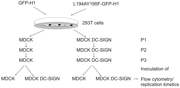
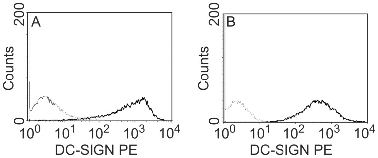
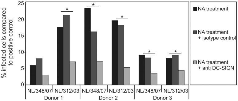
Dendritic cells express lectins receptors, like DC-SIGN, which allow these cells to sense glycans that are present on various bacterial and viral pathogens. Interaction of DC-SIGN with carbohydrate moieties induces maturation of dendritic cells and promotes endocytosis of pathogens which is an important property of these professional antigen presenting cells. Uptake of pathogens by dendritic cells may lead to cross-presentation of antigens or infection of these cells, which ultimately results in activation of virus-specific T cells in draining lymph nodes. Little is known about the interaction of DC-SIGN with influenza A viruses. Here we show that a virus with a non-functional receptor binding site in its hemagglutinin, can replicate in cells expressing DC-SIGN. Also in the absence of sialic acids, which is the receptor for influenza A viruses, these viruses replicate in DC-SIGN expressing cells including human dendritic cells. Furthermore, the efficiency of DC-SIGN mediated infection is dependent on the extent of glycosylation of the viral hemagglutinin.


Conflict of interest statement

Competing Interests: Two of the authors (AO and GR) also are employed by a company Viroclinics Biosciences BV. This does not alter the authors' adherence to all the PLOS ONE policies on sharing data and materials.

Figures

Figure 1
Figure 1. Schematic representation of the passage history and infection experiments with influenza viruses GFP-H1 and L194AY195F-GFP-H1.
Figure 2
Figure 2. DC-SIGN expression in stably transfected MDCK DC-SIGN and Vero DC-SIGN cells.
MDCK cells (A) and Vero cells (B) without (dotted line) and transfected with the gene encoding DC-SIGN (solid line) were analyzed for DC-SIGN expression after staining with a PE-labeled antibody to DC-SIGN and flow cytometry.
Figure 3
Figure 3. Replication kinetics of viruses GFP-H1 and L194AY195F-GFP-H1 in MDCK and DC-SIGN-expressing MDCK cells.
After transfection of 293T cells with reverse genetics plasmids, culture supernatants of influenza viruses GFP-H1 (A, C, E, G) and L194AY195F-GFP-H1 (B, D, F, H) virus passaged in MDCK (A–D) and MDCK-DC-SIGN (E–H) cells were obtained and used to inoculate MDCK (solid symbols) or MDCK DC-SIGN cells (open symbols) at a moi of 0.01. At the indicated time points post inoculation culture supernatant were tested for the infectious virus titers to determine the replication kinetics. Virus L194AY195F-GFP-H1 could not be rescued in MDCK cells.
Figure 4
Figure 4. GFP expression after infection with L194AY195F-GFP and GFP-H1 in MDCK and DC-SIGN-expressing MDCK cells.
MDCK and MDCK DC-SIGN cells were inoculated with influenza viruses GFP-H1 (A) and L194AY195F-GFP-H1 (B) at a MOI of 0.01 (solid lines). Both viruses were passaged in MDCK-DC-SIGN cells two or three times as indicated. Twenty-four hours post inoculation the cells were tested for GFP expression by flow cytometry. Uninfected cells were included as negative controls (dotted lines). Infection experiments with GFP-H1 virus passaged in MDCK cells essentially gave the same results as the virus passaged in MDCK DC-SIGN cells (data not shown).
Figure 5
Figure 5. Expression of DC-SIGN supports replication of influenza A viruses in the absence of sialic acids.
MDCK (A and C) and Vero cells (B and D) transfected with the DC-SIGN gene (black bars) or not (white bars), were treated with neuraminidase from vibrio cholerae and GolgiStop for 30 minutes to remove sialic acids from the cell surface. These cells were subsequently inoculated with five different A/H1N1 viruses (A and B) and four A/H3N2 viruses (C and D). The percentage infected cells relative to the untreated control cells, still possessing sialic acid, was assessed after detecting infected cells using a FITC-labelled antibody to the viral nucleoprotein and flow-cytometry. To confirm that the entry was mediated via DC-SIGN, Vero and Vero DC-SIGN were treated with neuraminidase from vibrio cholerae for 30 minutes to remove sialic acids from the cell surface and incubated with or without antibodies to DC-SIGN or an isotype control antibody as indicated (E and F). These cells were subsequently inoculated with influenza viruses. NL/312/03 and USSR/90/77. The percentage of infected cells compared to the positive control (untreated cells, still possessing sialic acid) was assessed as described above.
Figure 6
Figure 6. DC-SIGN expression on DC supports replication of influenza virus in absence of sialic acids.
DC were treated with neuraminidase from vibrio cholerae for 30 minutes to remove sialic acids from the cell surface and incubated with or without antibodies to DC-SIGN or an isotype control antibody as indicated. These cells were subsequently inoculated with two A/H3N2 influenza viruses. The percentage of infected cells compared to the positive control (untreated cells, still possessing of sialic acid) was assessed as described above.
Figure 7
Figure 7. The number of glycosylation sites present on HA determines the virus infection rates in DC-SIGN expressing cells.
MDCK (A) and Vero (B) cells, transfected with the DC-SIGN gene (black bars) or not (white bars) were treated with neuraminidase from vibrio cholerae and GolgiStop for 30 minutes to remove sialic acids from the cell surface. These cells were subsequently inoculated with A/Netherlands/26/07, A/Netherlands/26/07-Δ125, A/Netherlands/602/09, A/Netherlands/602/09-Δ276 or A/Netherlands/602/09-VN54 N125 N160. The percentage of infected cells relative to the positive control (untreated cells still possessing sialic acid) was assessed after detecting infected cells using a FITC-labeled antibody to the viral nucleoprotein and flow-cytometry.

References

    1. Svajger U, Anderluh M, Jeras M, Obermajer N (2010) C-type lectin DC-SIGN: an adhesion, signalling and antigen-uptake molecule that guides dendritic cells in immunity. Cell Signal 22: 1397–1405. - PMC - PubMed
    1. Geijtenbeek TB, Kwon DS, Torensma R, van Vliet SJ, van Duijnhoven GC, et al. (2000) DC-SIGN, a dendritic cell-specific HIV-1-binding protein that enhances trans-infection of T cells. Cell 100: 587–597. - PubMed
    1. Feinberg H, Mitchell DA, Drickamer K, Weis WI (2001) Structural basis for selective recognition of oligosaccharides by DC-SIGN and DC-SIGNR. Science 294: 2163–2166. - PubMed
    1. Yang ZY, Huang Y, Ganesh L, Leung K, Kong WP, et al. (2004) pH-dependent entry of severe acute respiratory syndrome coronavirus is mediated by the spike glycoprotein and enhanced by dendritic cell transfer through DC-SIGN. J Virol 78: 5642–5650. - PMC - PubMed
    1. Marzi A, Gramberg T, Simmons G, Moller P, Rennekamp AJ, et al. (2004) DC-SIGN and DC-SIGNR interact with the glycoprotein of Marburg virus and the S protein of severe acute respiratory syndrome coronavirus. J Virol 78: 12090–12095. - PMC - PubMed

Substances

LinkOut - more resources