. 2013 Feb;8(2):430-7.
doi: 10.1038/nprot.2012.153.

Production of hepatocyte-like cells from human pluripotent stem cells

Affiliations

Production of hepatocyte-like cells from human pluripotent stem cells

Nicholas R F Hannan et al. Nat Protoc. 2013 Feb.

Abstract

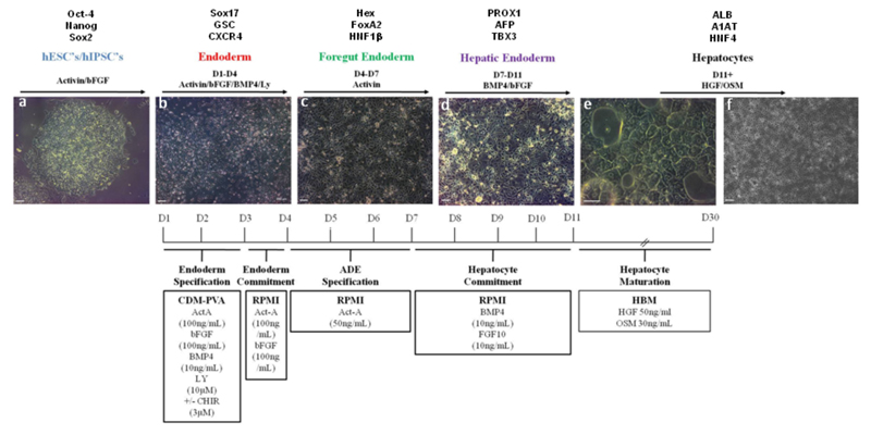
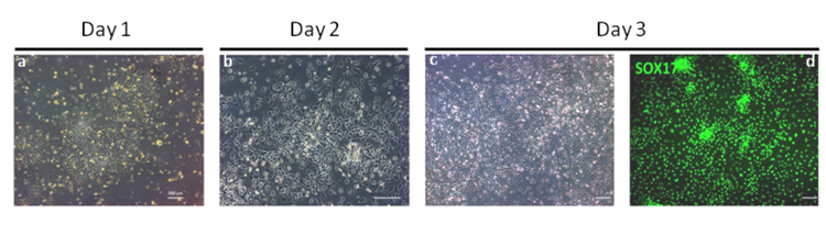
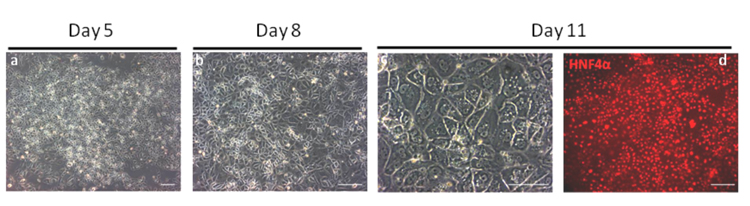
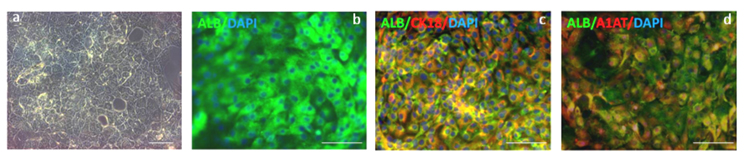
Large-scale production of hepatocytes from a variety of genetic backgrounds would be beneficial for drug screening and to provide a source of cells to be used as a substitute for liver transplantation. However, fully functional primary hepatocytes remain difficult to expand in vitro, and circumventing this problem by using an alternative source of cells is desirable. Here we describe a 25-d protocol to direct the differentiation of human pluripotent stem cells into a near-homogenous population of hepatocyte-like cells. As cells progress through this protocol, they express genes in a chronological manner similar to that described during in vivo hepatic development. The protocol relies on culture systems devoid of serum, feeders or complex extracellular matrices, which enable molecular analyses without interference from unknown factors. This approach works efficiently with human embryonic stem cells and human induced pluripotent stem cells and was recently used to model liver diseases in vitro.


Figures

Figure 1
Figure 1. Protocol to differentiate hPSCs into hepatocytes
Bright field pictures showing differentiation of hPSCs (H9, WiCell) into hepatocytes following a natural path of development. Panel a: Pluripotent cells are organised in tightly packed colonies with well-defined borders and display a high nuclear to cytoplasmic ratio. Panel b: Definitive endoderm specification starts with the migration of cells away from the original colony and with an increase in cell size. Panel c: Foregut endoderm cells have a polygonal / cobblestone shape while being organised into an epithelium. Panel d: Hepatic endoderm display canaliculi-like structures with a dark cytoplasm. As cells differentiate further, lipid vesicles can be observed and multi-nucleated cells appear. Panel e and f: hepatocyte-like cells generated after 25 days of differentiation . Differentiation time line detailing developmental stages with expected gene expression, basal medium and growth factor combinations and concentrations are provided (Text above and below panels). Scale bar: 200 μm.
Figure 2
Figure 2. Microphotographs showing endoderm differentiation of hPSCs
This stage is the most critical of the differentiation protocol as the production of homogenous population of DE cells is necessary for the efficient production of artificial hepatocytes. At Day 1 hESCs begin to undergo EMT marked by migration of cells away from the pluripotent colonies and start to express the primitive streak markers T, MIXL1 and EOMES. By Day 2 significant migration of cells has occurred and the original colony is no longer visible. The expression of T, and MIXL1 is downregulated while the expression of SOX17 and CXCR4 is induced. Panel a and b are bright field images of cell morphology on days 1 and 2 respectively. Panel c and d: By Day 3, endoderm cells homogenously express SOX17 (Panel c, bright field image; Panel d, Antibody: R&D AF1924) as shown by immunostaining. Under optimal conditions >90% of cells are CXCR4+/SOX17+ by FACS analyses. Scale bar: 200 μm.
Figure 3
Figure 3. Microphotographs showing Hepatic Specification
By day 5 of the protocol, differentiated cells starts mesenchymal to epithelium transition and express anterior foregut marker such as Sox17, FOXA2 and HHEX. At day 8, cells form a monolayer and express FOXA1, FOXA2 and HHEX. Panel a and b are bright field images of cell morphology on days 5 and 8 respectively. Panel c and d: At Day 11, hepatic cells display a specific appearance including a dark cytoplasm (Panel c, bright field image), light nucleus, and canaliculated borders. These cells express homogenously hepatic markers such as HNF4a (Panel d, Antibody SantaCruz sc6556) as shown by immunsotaining. Under optimal conditions >90% of cells are HNF4+ by FACS analyses. Scale bar: 200 μm.
Figure 4
Figure 4. Hepatocytes like cells generated after 20 days of differentiation
Cells expressing markers specific to hepatocytes such as albumin and α-1-antitrypsin are obtained after 20 days of differentiation. Panel a: The resulting cells display a distinct morphology common to primary human hepatocytes including multinuclei, distinct canaliculated border, and polygonal appearance. Panel b-d: Immunostaining analyses showing that under optimal conditions >85% ALB+/A1AT+ cells can be obtained (Panel b: immunostaining for Albumin (green; antibody R&D 188835); Panel c, co-immunostaining for Alb (green; antibody R&D 188835) and CK18 (red; antibody ab82254) Panel d, co-immunostaining for Alb (green; antibody R&D 188835) and A1AT(red; antibody DAKO A0012)). Panels b,c and d are all also counterstained with DAPI (blue) to show nuclei. Scale bar: 200 μm.

References

    1. Fisher RA, Strom SC. Human hepatocyte transplantation: worldwide results. Transplantation. 2006;82:441–449. - PubMed
    1. Thomson JA, et al. Embryonic stem cell lines derived from human blastocysts. Science. 1998;282:1145–1147. - PubMed
    1. Takahashi K, et al. Induction of pluripotent stem cells from adult human fibroblasts by defined factors. Cell. 2007;131:861–872. - PubMed
    1. Rashid ST, Vallier L. Induced pluripotent stem cells--alchemist’s tale or clinical reality? Expert Rev Mol Med. 2010;12:25. - PubMed
    1. Rashid ST, et al. Modeling inherited metabolic disorders of the liver using human induced pluripotent stem cells. J Clin Invest. 2010;120:3127–3136. - PMC - PubMed

Publication types