. 2013 Apr;222(4):473-80.
doi: 10.1111/joa.12030. Epub 2013 Feb 21.

Expression of orexin A and its receptor 1 in the human prostate

Affiliations

Expression of orexin A and its receptor 1 in the human prostate

Salvatore Valiante et al. J Anat. 2013 Apr.

Abstract

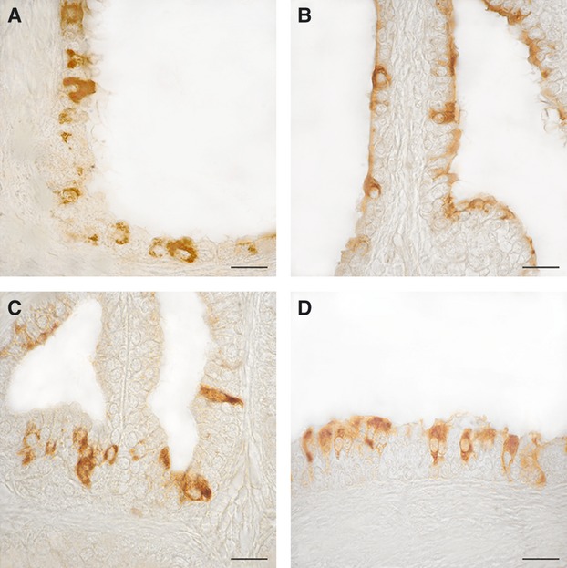
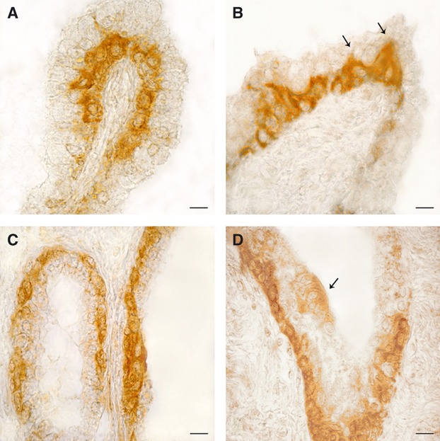
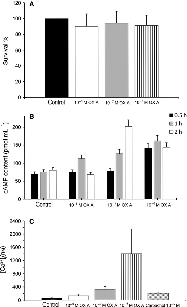
The peptides orexin A (OXA) and orexin B, deriving from the cleavage of the precursor molecule prepro-orexin, bind two G-coupled transmembrane receptors, named as receptor 1 (OX1R) and receptor 2 for orexin, showing different affinity-binding properties. First discovered in the rat hypothalamus, orexins and their receptors have been also found in many peripheral tissues where they exert neuroendocrine, autocrine and paracrine functions. Because inconclusive data on their localization in the mammalian prostate are reported, the aim of this study was to investigate the presence of prepro-orexin, OXA and OX1R in the human normal and hyperplastic gland. Immunohistochemistry revealed the localization of both OXA and OX1R in the cytoplasm of the follicular exocrine epithelium of all tested normal and hyperplastic prostates. Positive immunostaining was mainly observed in the basal cells of the stratified epithelium, and only rarely in the apical cells. The expression of mRNAs coding for prepro-orexin and OX1R and of proteins in the tissues was also ascertained by polymerase chain reaction and Western blotting analysis, respectively. In order to gain insights into the functional activity of OXA in the prostate, we administered different concentrations of OXA to cultured prostatic epithelial cells PNT1A. We first demonstrated that PNT1A cells express OX1R. The addition of OXA did not affect PNT1A cell proliferation, while it enhanced cAMP synthesis and Ca(2+) release from intracellular storage. Overall, our results definitely demonstrate the expression of OXA and OX1R in the human prostate, and suggest an active role for them in the metabolism of the gland.


Figures

Fig. 1
Fig. 1
OXA- (A and B) and OX1R- (C and D) immunoreactivity in the human normal prostate. (A) Positive cells scattered along the monolayered epithelium of a prostatic follicle. (B) The majority of positive cells visible in this micrograph contain differently stained granular material in the apical portion of their cytoplasm. (C and D) Positive cells lined along the stratified epithelium of three follicles. Some of them are elongated in shape and completely filled with clusters of immunoreactive granules. Avidin–biotin immunohistochemical method. Scale bars: 20 μm.
Fig. 2
Fig. 2
OXA- (A and B) and OX1R- (C and D) immunoreactivity in the human hyperplastic prostate. (A) The basal membrane of an intrafollicular, papillar-like structure is lined by a continuous row of intensely stained cells. (B) A peculiarity often shown by positive basal cells is the presence of a slender cytoplasmic extension (arrows) directed towards the follicular lumen and intermingled between the negative apical cells. (C) Almost all basal cells of the follicular epithelium contain OX1R-immunoreactive material, which completely fills their cytoplasm. (D) Invagination of a prostatic follicle completely lined by positive basal cells. The arrow points to a small cluster of low intensely stained apical cells close in contact with the follicular fluid. Avidin–biotin immunohistochemical method. Scale bars: 20 μm.
Fig. 3
Fig. 3
Expression of prepro-orexin and OX1R mRNAs and the proteins in human normal and hyperplastic prostates (A and B) and expression of OX1R in PNT1A cultured cells (C). (A) RT-PCR analysis. Lane 1, DNA ladder; lane 2, prepro-orexin and OX1R mRNA transcripts from whole rat brain (positive control); lanes 3 and 4, prepro-orexin and OX1R mRNA transcripts from normal and hyperplastic prostate samples, respectively; lane 5, negative control (no cDNA input). The bottom of (A) reports the beta-actin mRNA transcripts (internal control). (B) Western blotting analysis. Lane 1, homogenates from whole rat brain (positive control); lanes 2 and 3, homogenates from normal and hyperplastic prostate tissue, respectively; lane 4, prostate homogenates treated with the antisera directed against prepro-orexin or OX1R pre-absorbed with their respective control peptides (negative control). (C) Western blotting analysis. Lane 1, homogenates from whole rat brain (positive control); lane 2, homogenates from PNT1A cell lysate; lane 3, cell lysates treated with the antiserum directed against OX1R pre-absorbed with its control peptide (negative control). The upper blots of (B) and (C) were stripped and re-probed with an anti-tubulin monoclonal antibody to ensure equal loading of proteins in all lanes (lower blots). Molecular mass markers are indicated on the left of the Western blotting panels. Similar results were obtained from four separate experiments of identical design.
Fig. 4
Fig. 4
Effects of different OXA concentrations on PNT1A cells. (A) Results from MTT-based cell proliferation assay. The data, expressed as mean ± SEM of four independent experiments, are reported as the survival percentage as compared with control cells, after 24 h of OXA administration. (B) cAMP production induced by OXA at different time intervals. Data are expressed as mean ± SEM of three independent experiments: 0.5 h 10−6 m compared with control, P < 0.05; 1 h each dose compared with control, P < 0.05, 2 h 10−7, 10−6 m compared with control, P < 0.05. (C) Intracellular Ca2+ release induced by different OXA doses. Control was calcium-free medium exclusively, other samples were calcium-free. Carbachol 10−6 m was used as positive control. Data are expressed as mean ± SEM of three independent experiments. All experimental groups have a value of P < 0.05 compared with control.

References

    1. Ammoun S, Holmqvist T, Shariatmadari R, et al. Distinct recognition of OX1 and OX2 receptors by orexin peptides. J Pharmacol Exp Ther. 2003;305:507–514. - PubMed
    1. Assisi L, Tafuri S, Liguori G, et al. Expression and role of receptor 1 for orexins in seminiferous tubules of rat testis. Cell Tissue Res. 2012;348:601–607. - PubMed
    1. Barreiro ML, Pineda R, Navarro VM, et al. Orexin 1 receptor messenger ribonucleic acid expression and stimulation of testosterone secretion by orexin-A in rat testis. Endocrinology. 2004;145:2297–2306. - PubMed
    1. Barreiro ML, Pineda R, Gaytan F, et al. Pattern of orexin expression and direct biological actions of orexin-A in rat testis. Endocrinology. 2005;146:5164–5175. - PubMed
    1. Blanco M, García-Caballero T, Fraga M, et al. Cellular localization of orexin receptors in human adrenal gland, adrenocortical adenomas and pheochromocytomas. Regul Pept. 2002;104:161–165. - PubMed

Publication types

LinkOut - more resources