. 2013 Mar;123(3):1241-61.
doi: 10.1172/JCI61271. Epub 2013 Feb 15.

Cigarette smoke mediates epigenetic repression of miR-487b during pulmonary carcinogenesis

Affiliations

Cigarette smoke mediates epigenetic repression of miR-487b during pulmonary carcinogenesis

Sichuan Xi et al. J Clin Invest. 2013 Mar.

Abstract

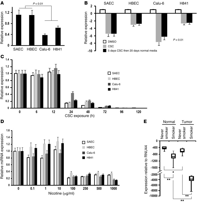
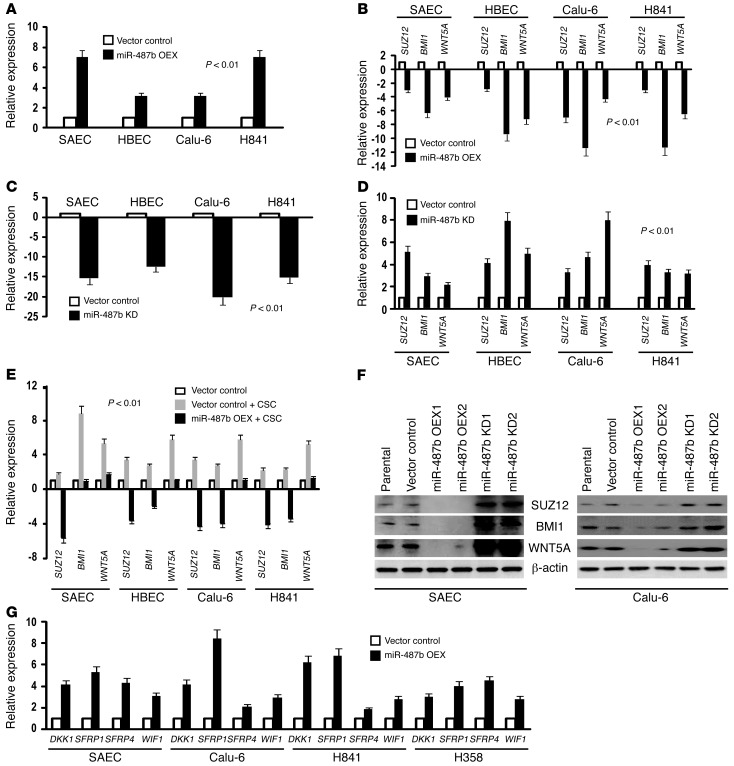
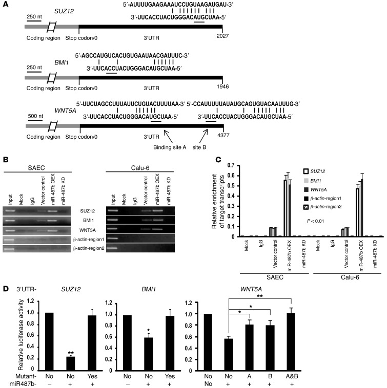
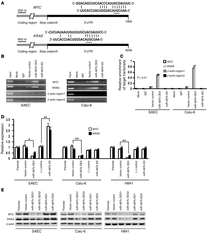
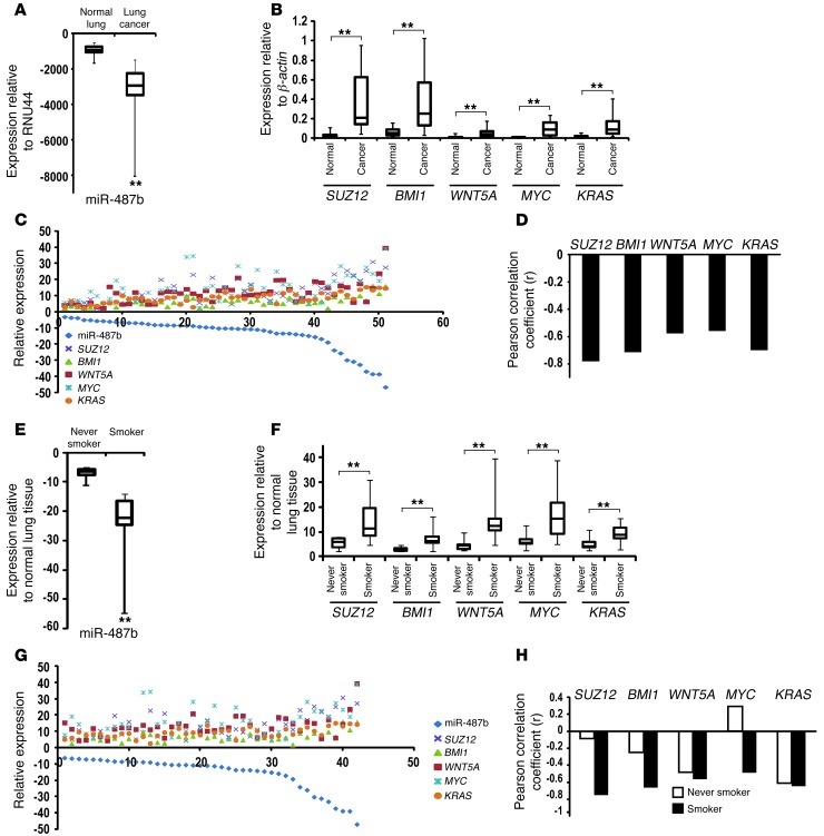
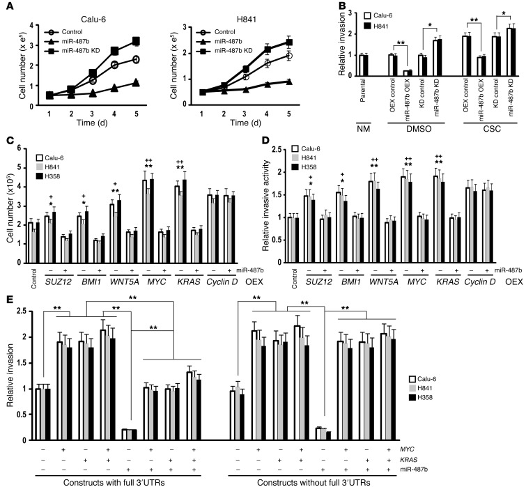
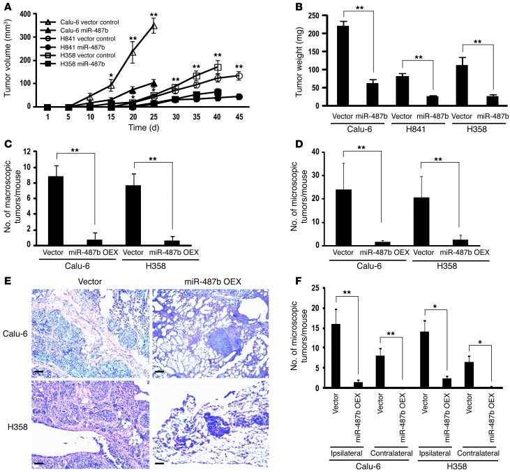
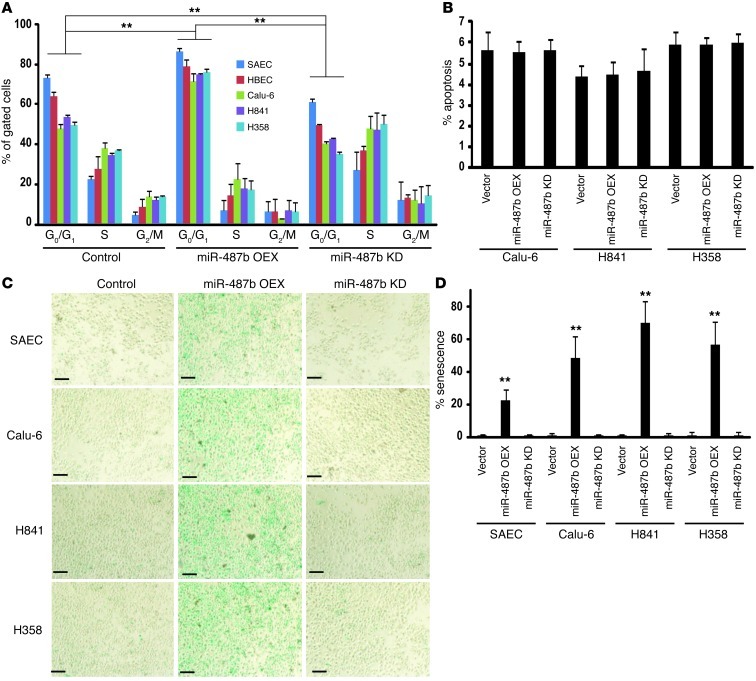
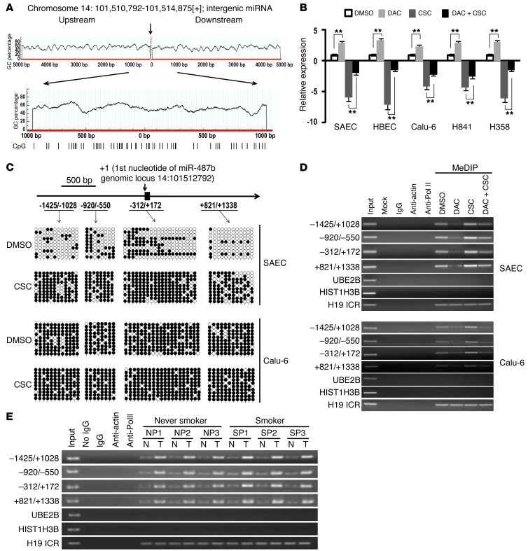
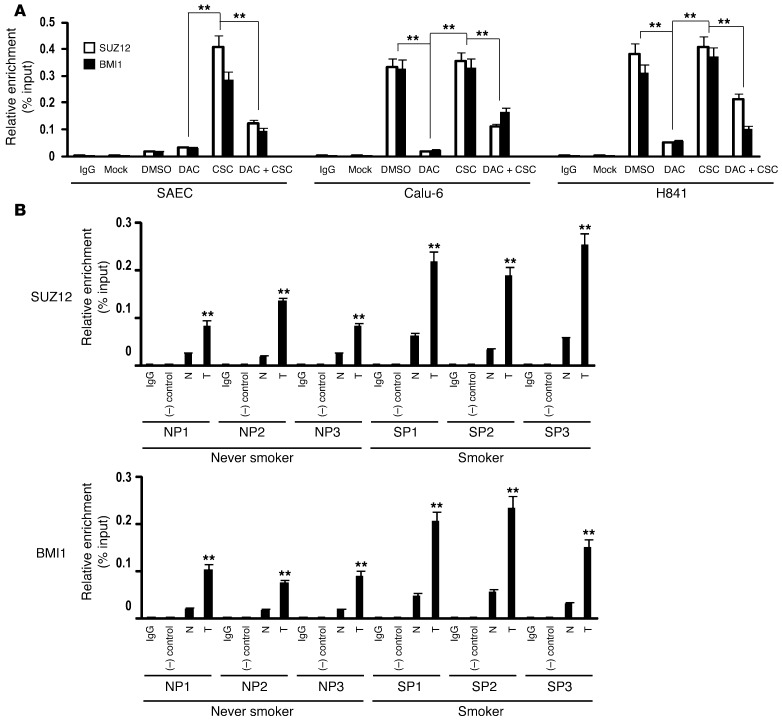
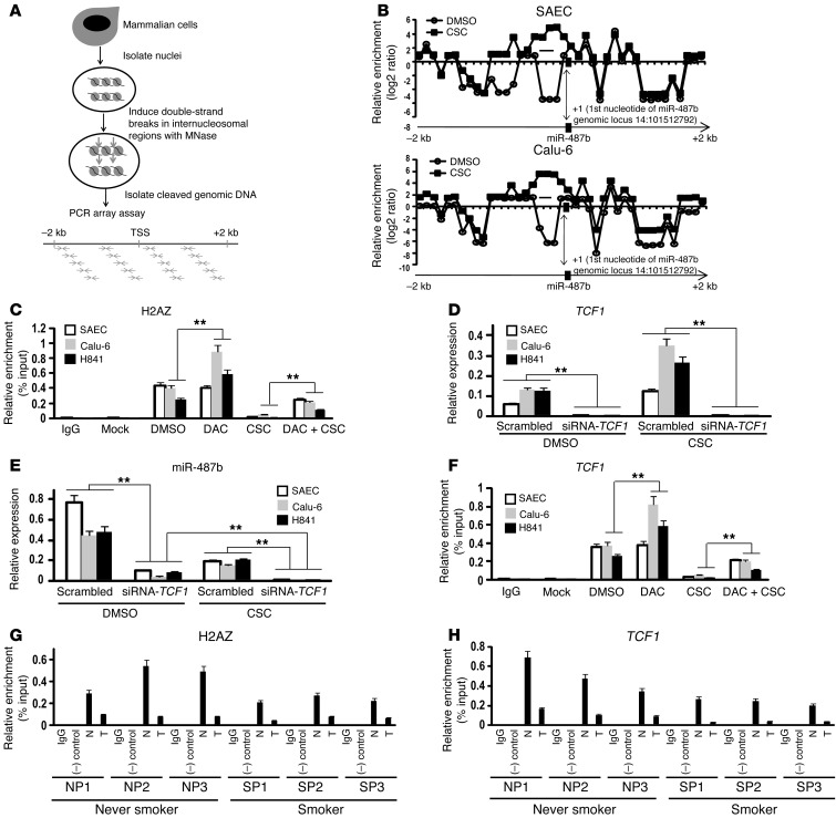
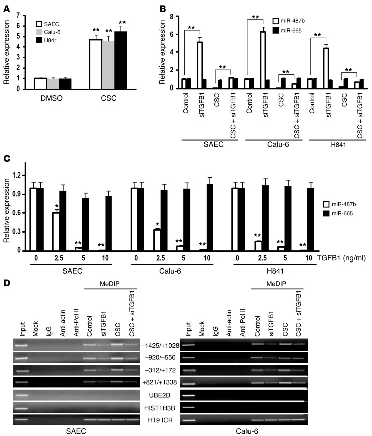
MicroRNAs are critical mediators of stem cell pluripotency, differentiation, and malignancy. Limited information exists regarding microRNA alterations that facilitate initiation and progression of human lung cancers. In this study, array techniques were used to evaluate microRNA expression in normal human respiratory epithelia and lung cancer cells cultured in the presence or absence of cigarette smoke condensate (CSC). Under relevant exposure conditions, CSC significantly repressed miR-487b. Subsequent experiments demonstrated that miR-487b directly targeted SUZ12, BMI1, WNT5A, MYC, and KRAS. Repression of miR-487b correlated with overexpression of these targets in primary lung cancers and coincided with DNA methylation, de novo nucleosome occupancy, and decreased H2AZ and TCF1 levels within the miR-487b genomic locus. Deoxy-azacytidine derepressed miR-487b and attenuated CSC-mediated silencing of miR-487b. Constitutive expression of miR-487b abrogated Wnt signaling, inhibited in vitro proliferation and invasion of lung cancer cells mediated by CSC or overexpression of miR-487b targets, and decreased growth and metastatic potential of lung cancer cells in vivo. Collectively, these findings indicate that miR-487b is a tumor suppressor microRNA silenced by epigenetic mechanisms during tobacco-induced pulmonary carcinogenesis and suggest that DNA demethylating agents may be useful for activating miR-487b for lung cancer therapy.


Figures

Figure 1
Figure 1. Expression of miR-487b in cultured cells as well as primary lung cancers and adjacent normal lung tissues.
(A) qRT-PCR analysis of endogenous miR-487b expression normalized with control miRNA (RNU44) in SAECs, HBECs, Calu-6, and H841 cells. (B) qRT-PCR analysis demonstrating downregulation of miR-487b in cultured cells following 5-day CSC exposure. Repression of miR-487b persisted following discontinuation of CSC treatment. (C) qRT-PCR analysis demonstrating time-dependent repression of miR-487b in SAECs, HBECs, Calu-6, and H841 cells following CSC exposure. (D) qRT-PCR analysis demonstrating dose-dependent repression of miR-487b in lung cancer cells by nicotine, which leveled off with concentrations of 100 μg/ml or more following 5-day exposure. (E) qRT-PCR analysis of miR-487b expression in human lung cancers relative to paired adjacent normal lung tissues (n = 51). miR-487b levels were significantly lower in cancers relative to corresponding adjacent normal lung tissues. Furthermore, miR-487b levels were significantly more repressed in lung cancers from active/former smokers compared with those from never smokers. *P < 0.05; **P < 0.01.
Figure 2
Figure 2. miR-487b negatively regulates SUZ12, BMI1, and WNT5A in cultured normal respiratory epithelial and lung cancer cells.
(A) qRT-PCR analysis demonstrating miR-487b expression in normal respiratory epithelia and lung cancer cells constitutively expressing miR-487b (miR-487b OEX) relative to vector controls. (B) qRT-PCR analysis demonstrating that overexpression of miR-487b downregulates SUZ12, BMI1, and WNT5A in normal respiratory epithelia and lung cancer cells. (C) qRT-PCR analysis demonstrating decreased levels of endogenous miR-487b in SAECs, HBECs, Calu-6, and H841 cells following transient knockdown of miR-487b (miR-487b KD) relative to vector controls. (D) qRT-PCR analysis demonstrating that downregulation of miR-487b enhances levels of SUZ12, BMI1, and WNT5A expression in SAECs, HBECs, Calu-6, and H841 cells. (E) qRT-PCR analysis demonstrating that overexpression of miR-487b abrogates CSC-mediated increases in SUZ12, BMI1, and WNT5A in SAECs, HBECs, Calu-6, and H841 cells. (F) Immunoblot analysis of SUZ12, BMI1, and WNT5A expression in parental and vector control SAECs and Calu-6 cells, as well as SAECs and Calu-6 cells exhibiting constitutive overexpression or knockdown of miR-487b. Results of immunoblot experiments were consistent with qRT-PCR analysis (B and D). (G) Results of qRT-PCR analysis demonstrating that overexpression of miR-487b upregulates Wnt antagonists DKK1, SFRP1, SFRP4, and WIF1 in normal SAECs as well as Calu-6, H841, and H358 lung cancer cells.
Figure 3
Figure 3. SUZ12, BMI1, and WNT5A are direct targets of miR-487b.
(A) Putative target sites of miR-487b within SUZ12, BMI1, and WNT5A 3′ UTRs (top, middle, and bottom, respectively). (B and C) Ago-CLIP (B) and quantitative Ago-CLIP (C) analyses of interactions of miR-487b with SUZ12, BMI1, and WNT5A 3′ UTRs in SAECs and Calu-6 cells. Overexpression of miR-487b significantly increased precipitation of SUZ12, BMI1, and WNT5A, indicating direct interaction of miR-487b with the respective 3′ UTRs. (D) Luciferase reporter assays demonstrating that overexpression of miR-487b decreases luciferase activities of WT but not mutant 3′ UTRs of SUZ12, BMI1, and WNT5A in SAECs. For WNT5A, miR-487b–binding sites A and/or B were mutated; mutation of both miR-487b binding sites was more effective than single mutations in abrogating the effect of miR-487b. *P < 0.05; **P < 0.01.
Figure 4
Figure 4. miR-487b targets MYC and KRAS.
(A) Putative binding sites of miR-487b within MYC and KRAS 3′ UTRs (upper and lower panels, respectively). (B and C) Ago-CLIP (B) and quantitative Ago-CLIP (C) analyses depicting direct interaction of miR-487b with 3′ UTRs of MYC and KRAS in SAECs and Calu-6 cells. (D) qRT-PCR analysis of MYC and KRAS expression in parental SAECs, Calu-6, or H841 cells, vector controls, or corresponding cells exhibiting overexpression or knockdown of miR-487b. *P < 0.05; **P < 0.01. (E) Immunoblot analysis of MYC and KRAS expression in SAECs and Calu-6 and H841 cells following overexpression or knockdown of miR-487b. Overexpression of miR-487b decreased MYC and KRAS levels in all 3 lines. Knockdown of miR-487b increased MYC and KRAS levels above that of controls in SAECs and, to a lesser extent, in Calu-6 and H841 cells.
Figure 5
Figure 5. Reverse correlation between miR-487b and its 5 targets relative to smoking history of patients.
(A) qRT-PCR analysis of miR-487b expression in 51 lung cancer specimens relative to paired adjacent normal lung tissues. (B) qRT-PCR analysis demonstrating increased SUZ12, BMI1, WNT5A, MYC, and KRAS expression levels in lung cancers relative to paired adjacent normal lung tissues. (C and D) Correlation patterns (C) and corresponding correlation coefficient values (D) between miR-487b and its targets in tumor specimens relative to paired adjacent normal lung tissues (n = 51). (E) qRT-PCR analysis of miR-487b expression in lung cancers from smokers/former smokers versus never smokers. (F) qRT-PCR analysis demonstrating increased SUZ12, BMI1, WNT5A, MYC, and KRAS expression levels in lung cancers from smokers/former smokers relative to never smokers. (G) Correlation patterns between miR-487b and its targets in 42 lung cancers from smokers. (H) Corresponding coefficient values between miR-487b repression and upregulation of 5 targets in 9 lung cancers from never smokers and 42 lung cancers from smokers/former smokers. **P < 0.01.
Figure 6
Figure 6. miR-487b functions as a tumor suppressor in lung cancer cells.
(A) Effects of miR-487b expression on in vitro proliferation of Calu-6 and H841 lung cancer cells. (B) Matrigel invasion assays demonstrating that overexpression of miR-487b inhibited, whereas knockdown of miR-487b enhanced, invasion of Calu-6 and H841 lung cancer cells. miR-487b significantly inhibited CSC-induced invasion of lung cancer cells. The effects of miR-487b knockdown on invasion of DMSO-treated cells were comparable to these observed in vector controls exposed to CSC. *P < 0.05; **P < 0.01. (C) Cell count assays demonstrating effects of miR-487b on proliferation of lung cancer cells overexpressing SUZ12, BMI1, WNT5A, MYC, or KRAS. miR-487b had no effects on proliferation induced by cyclin D1, which is not targeted by miR-487b. +P < 0.05; ++P < 0.01 vs. control. *P < 0.05; **P < 0.01 vs. miR-487b. (D) Matrigel assays demonstrating effects of miR-487b on invasive potential of lung cancer cells following overexpression of the 5 miR-487 targets. miR-487b had no effects on invasion induced by cyclin D1. +P < 0.05; ++P < 0.01 vs. control. *P < 0.05; **P < 0.01 vs miR-487b. (E) Specificity of effects of miR-487b expression on invasion potential of lung cancer cells mediated by MYC and KRAS. miR-487b failed to inhibit invasion induced by overexpression of MYC and KRAS lacking full 3′ UTR sequences.
Figure 7
Figure 7. Effects of miR-487b on growth and metastatic potential of lung cancer cells in vivo.
(A) Growth of Calu-6, H841, and H358 subcutaneous xenografts in nude mice. Volumes of xenografts derived from Calu-6, H841, or H358 cells overexpressing miR-487b were significantly smaller than control xenografts. (B) Tumor masses from Calu-6, H841, and H358 xenografts. miR-487b overexpression significantly decreased average mass of tumor xenografts. (C) Quantitation of macroscopic pulmonary nodules derived from Calu-6 or H358 cells stably expressing miR-487b or control vectors. (D) Quantitation of total microscopic pulmonary tumors following orthotopic injection of Calu-6 or H358 cells stably expressing miR-487b or control vectors. (E) Representative microscopic images (H&E staining) of intrapulmonary tumors from Calu-6 or H358 cells stably expressing miR-487b or control vectors. Expression of miR-487b decreased invasion of lung cancer cells in vivo. Scale bars: 50 mm. Original magnification, ×100. (F) Quantitation of ipsilateral and contralateral microscopic pulmonary tumors following orthotopic injection of Calu-6 or H358 cells stably expressing miR-487b or control vectors. *P < 0.05; **P < 0.01.
Figure 8
Figure 8. miR-487b inhibits cellular proliferation via cell-cycle arrest and senescence but not apoptosis in normal respiratory epithelia and lung cancer cells.
(A) Flow cytometry analysis demonstrating that miR-487b overexpression significantly impairs progression of SAECs, HBECs, Calu-6, H841, and H358 cells from G0/G1 into S and G2/M phases. Endogenous depletion of miR-487b enhanced cell-cycle progression in these cells. Data are represented as percentage of gated cells ± SD. (B) In vitro apoptosis assay demonstrating no change in apoptosis index in lung cancer cells exhibiting overexpression or knockdown of miR-487b. (C) Representative microscopic images (senescence staining) of SAECs, Calu-6, H841, and H358 cells exhibiting overexpression or depletion of miR-487b. Scale bars: 50 mm. Original magnification, ×100. (D) Quantitation of miR-487b–induced cellular senescence in normal respiratory epithelia and lung cancer cells. Ectopic expression of miR-487b significantly increased percentage of senescence in these cells. However, depletion of miR-487b did not affect senescence in these cells. Data are represented as percentage of cells ± SD. **P < 0.01.
Figure 9
Figure 9. Epigenetic alterations coinciding with CSC-mediated repression of miR-487b.
(A) Schematic depiction demonstrating higher CpG density 5 kb up- and downstream of the miR-487b genomic locus ( http://www.urogene.org/methprimer/). (B) qRT-PCR analysis demonstrating that DAC significantly increases expression of primary miR-487b and attenuates CSC-mediated repression of miR-487b in SAECs, HBECs, Calu-6, H841, and H358 cells. **P < 0.01. (C) Bisulfite sequencing of 4 genomic regions around the miR-487b genomic locus, demonstrating that CSC enhances DNA methylation in SAECs. CSC did not alter CpG methylation around miR-487b genomic locus in Calu-6 cells exhibiting higher pretreatment DNA methylation within these genomic sites. (D) MeDIP analysis of DNA methylation profiles within the miR-487b genomic locus in SAECs and Calu-6 cells treated with DAC or CSC. DAC significantly decreased methylation near miR-487b in SAECs and Calu-6 cells and partially abrogated CSC-mediated CpG methylation in these cells. (E) MeDIP analysis of DNA methylation profiles within the miR-487b genomic locus in human lung cancers relative to paired adjacent normal lung tissues. CpG methylation levels near miR-487b were higher in tumors relative to corresponding adjacent normal lung tissues. Furthermore, methylation levels in these regions appeared higher in lung cancers from active/former smokers compared with those from never smokers.
Figure 10
Figure 10. SUZ12 and BMI1 are involved in silencing miR-487b.
(A) Quantitative ChIP analysis of SUZ12 and BMI1 levels within the miR-487b genomic locus in SAECs and Calu-6 and H841 cells. DAC decreased occupancy of SUZ12 and BMI1 in this region in lung cancer cells and significantly attenuated CSC-mediated recruitment of these polycomb proteins to the miR-487b–regulating region. (B) Quantitative ChIP analysis of the miR-487b genomic locus in human lung cancers relative to paired adjacent normal lung tissues. Levels of SUZ12 and BMI1 within the miR-487b genomic locus in tumors (T) were significantly higher than corresponding adjacent normal (N) lung tissues. Furthermore, SUZ12 and BMI1 levels were higher in lung cancers from active/former smokers compared with those from never smokers. N vs. T for each patient: P < 0.01; never smoker vs. smoker for both N and T: P < 0.05. **P < 0.01.
Figure 11
Figure 11. CSC-induced chromatin remodeling coincides with miR-487b transcription.
(A) Sketch diagram for MNase protection assay. (B) Results of MNase protection assays depicting 2 upstream NFR (+50 to +350 bp and +650 to +900 bp) of miR-487b in control SAECs and 1 NFR (+50 to +350) in untreated Calu-6 cells. CSC induced de novo nucleosome occupancy of these regions. (C) Quantitative ChIP analysis depicting H2AZ levels in the first NFR of miR-487b. DAC partially attenuated CSC-mediated decreases in H2AZ levels. See text for details. (D and E) qRT-PCR analysis demonstrating that CSC induces TCF1 expression in SAECs and Calu-6 and H841 cells (D); knockdown of TCF1 inhibits basal expression of miR-487b in SAECs and Calu-6 and H841 cells and augments CSC-mediated repression of miR-487b in these cells (E). (F) Quantitative ChIP analysis demonstrating decreased occupancy of TCF1 in the first NFR of miR-487b in SAECs and Calu-6 and H841 cells following CSC exposure. DAC increased TCF1 levels in this NFR in Calu-6 and H841 cells and attenuated CSC-mediated decreases in TCF1 occupancy in this NFR in SAECs and Calu-6 and H841 cells. *P < 0.05; **P < 0.01. (G, H) Quantitative ChIP analysis demonstrating significantly lower levels of H2AZ (G) and TCF1 (H) in the first NFR of miR-487b in human lung cancers relative to paired normal lung tissues. H2AZ and TCF1 enrichment levels were lower in lung cancers from active/former smokers compared with those from never smokers. N vs. T for each patient: P < 0.01; never smoker vs. smoker for both N and T: P < 0.05.
Figure 12
Figure 12. TGFB1 mediates repression of miR-487b by CSC.
(A) qRT-PCR analysis demonstrating that CSC enhances TGFB1 expression in SAECs and Calu-6 and H841 cells. (B) qRT-PCR analysis demonstrating that knockdown of TGFB1 markedly increases miR-487b expression and abrogates CSC-mediated repression of miR-487b in SAECs and to a lesser extent in Calu-6 and H841 cells. Knockdown of TGFB1 had no effect on miR-665 expression in these cells. (C) qRT-PCR analysis demonstrating dose-dependent decreases in miR-487b (but not miR-665) in SAECs and Calu-6 and H841 cells following 48-hour exposure to recombinant TGFB1. *P < 0.05; **P < 0.01. (D) MeDIP analysis demonstrating that knockdown of TGFB1 markedly decreases DNA methylation levels in untreated SAECs and Calu-6 cells and diminishes CSC-mediated increases in DNA methylation around the miR-487b genomic locus in these cells.

References

    1. Malumbres M. miRNAs and cancer: an epigenetics view [published online ahead of print July 4, 2012]. Mol Aspects Med. doi: 10.1016/j.mam.2012.06.005. doi: 10.1016/j.mam.2012.06.005 . - DOI - DOI - PMC - PubMed
    1. Jansson MD, Lund AH. MicroRNA and cancer. Mol Oncol. 2012;6(6):590–610. doi: 10.1016/j.molonc.2012.09.006. - DOI - PMC - PubMed
    1. Iorio MV, Croce CM. MicroRNA dysregulation in cancer: diagnostics, monitoring and therapeutics. A comprehensive review. EMBO Mol Med. 2012;4(3):143–159. doi: 10.1002/emmm.201100209. - DOI - PMC - PubMed
    1. Lee D, Shin C. MicroRNA-target interactions: new insights from genome-wide approaches. Ann N Y Acad Sci. 2012;1271:118–128. doi: 10.1111/j.1749-6632.2012.06745.x. - DOI - PMC - PubMed
    1. Zardo G, et al. Transcriptional targeting by microRNA-Polycomb complexes: A novel route in cell fate determination. Cell Cycle. 2012;11(19):3543–3549. doi: 10.4161/cc.21468. - DOI - PMC - PubMed

Publication types

MeSH terms

Substances