Review
. 2013 Jul 1;5(7):a013383.
doi: 10.1101/cshperspect.a013383.

Expanding proteostasis by membrane trafficking networks

Affiliations
Review

Expanding proteostasis by membrane trafficking networks

Darren M Hutt et al. Cold Spring Harb Perspect Biol. .

Abstract

The folding biology common to all three kingdoms of life (Archaea, Bacteria, and Eukarya) is proteostasis. The proteostasis network (PN) functions as a "cloud" to generate, protect, and degrade the proteome. Whereas microbes (Bacteria, Archaea) have a single compartment, Eukarya have numerous subcellular compartments. We examine evidence that Eukarya compartments use coat, tether, and fusion (CTF) membrane trafficking components to form an evolutionarily advanced arm of the PN that we refer to as the "trafficking PN" (TPN). We suggest that the TPN builds compartments by generating a mosaic of integrated cargo-specific trafficking signatures (TRaCKS). TRaCKS control the temporal and spatial features of protein-folding biology based on the Anfinsen principle that the local environment plays a critical role in managing protein structure. TPN-generated endomembrane compartments apply a "quinary" level of structural control to modify the secondary, tertiary, and quaternary structures defined by the primary polypeptide-chain sequence. The development of Anfinsen compartments provides a unifying foundation for understanding the purpose of endomembrane biology and its capacity to drive extant Eukarya function and diversity.


Figures

Figure 1.
Figure 1.
Proteostasis and trafficking biology. The hierarchical influence of proteostasis on proteome function (indicated by the dark orange circle with a protein represented by a black node) in a eukaryotic cell. The first layer outside the proteome lists the working components of the proteostasis network (PN; blue lettering) that we now propose includes membrane trafficking biology coat, tether, and fusion (CTF) components comprising the trafficking proteostasis network (TPN; see main text). The next layer lists the many signaling pathways (green lettering) that can influence the composition of the PN/TPN in each cell type. The outer layer (brown lettering) highlights the impact of genetics (including modifiers), epigenetics (including HATs and HDACs), metabolites, and physiological stress pathways stemming from the extracellular environment (solid green) that influence PN/TPN activity. We capture the dynamic features of these relationships as the “cloud” (light orange icon), a unique TPN/PN management system that surrounds each protein (black node) and controls its function from birth to death. (Adapted from Powers et al. 2009.)
Figure 2.
Figure 2.
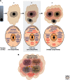
The contribution of compartmentalization to proteostasis biology. (A, upper panels) Illustrated is the relationship between protein (black sphere) folding in vitro (column 1) and biological protein folding in vivo (columns 2–4), the latter requiring the assistance of PN/TPN components (lower panels). In column 1, protein folding in vitro is limited to the chemical information contained in the polypeptide chain sequence and is strongly influenced by the choice of the folding buffer. In the simplest case in vivo, illustrated by column 2 (such as found in extant Bacteria and Archaea), one cytosolic PN (the orange cloud) manages all intracellular folding and the export of proteins to and through the cell surface, although specialized chaperones can manage folding in the environment immediately outside the cell (Evans et al. 2011; Powers and Balch 2011; Quan et al. 2011). The small gray cloud icon surrounding the protein (dark circle) defines the select PN components used by a particular protein to facilitate its own structure/function relationships. In column 3, the addition of an intracellular folding compartment (e.g., the ER found in eukaryotic cells) generates a specialized folding environment that now requires trafficking biology found in the cytosol (hazy red cloud) to facilitate cargo movement between the compartment and the cell surface. (Gray rectangle) Indicates that this is only an ancestral state found in the last universal common ancestor (LUCA). In column 4, the presence of multiple compartments (>2) found in extant eukaryotic cells is accompanied by the evolution of CTF-based trafficking pathways (hazy red cloud icon) found in the cytosol that manage both compartment identity and itinerant cargo flow between compartments. (B) Illustrated are prominent CTF-defined compartments that can affect the structure/function relationships of the protein fold in eukaryotic cells. The large orange cloud icon highlights cytosolic-localized PN. Trafficking components (hazy red cloud) that manage compartment composition, structure, and function are localized to the cytosol with the exception of the fusion machinery components that are often associated with the membrane. The dashed boundary around the ER (purple) highlights the fact that it can physically exchange content with the cytosol. The dashed boundary around the autophagic-phagocytic-lysosome (red) cloud icon highlights the ability of the autophagic systems to sample intra- and extracellular cargo directly.
Figure 3.
Figure 3.
The TPN and TRaCKS. (A) C, T, and F components work together to form the TPN (hazy red cloud icon). The TPN is unique for each cell type, and temporally and spatially generates endomembrane compartments (Gurkan et al. 2005). In response to cargo (black circle), select CTF components generate TRaCKS (dotted circle) that specifically manage the trajectory and function of cargo in maintaining or transiting through endomembrane compartments. The mosaic composition of TRaCKS (right panel) can be viewed as contributing to an additional “quinary” level of structural interactions to the secondary, tertiary, and quaternary structures defined by primary sequence of the polypeptide chain. (B) Speculative snapshot of a mosaic arrangement of TRaCKS used to generate compartment (dotted gray lines) identities or to facilitate flow between compartments based on the kinetic (K) and/or thermodynamic (T) properties of the cargo client interaction with PN and TPN components (K1T1, K2T2, …). A and B are compartment-generating TRaCKS (e.g., glycosyl transferases), and C is an itinerant TRaCKS (e.g., CFTR) permitting rapid transit through compartments (Stagg et al. 2007; Powers et al. 2009).
Figure 4.
Figure 4.
Management of glycan structure and neurotransmission by TRaCKS. (A) Snapshot of a specific collection of coat (purple oval), tether (green oval), or fusion (brown oval) components (ovals) contributing to the TRaCKS (dotted red line) mediating synaptic vesicle docking and fusion at the synapse. The activity of these TRaCKS components (hazy red cloud icon) are coupled to the activity of cytosolic Hsp40, Hsc/p70, and Hsp90 protein-folding components (orange cloud icon). (B) Snapshot of select CTF components (ovals) contributing to TRaCKS (dotted red circle) involved in glycosyl transferase localization (black circle) to medial-trans Golgi compartments as described in A. (C) Currently recognized “morphological” compartments (ER, Golgi, endosomes, etc.) are temporally and spatially managed by the TPN (hazy red cloud icon) to form an integrated endomembrane system (multicolored, overlapping cloud icons outlined by a solid gray line). In this view, the ER forms a central, multitasking proteostasis hub that is linked to most (if not all) endomembrane trafficking pathways through the TPN—including mitochondria and peroxisomes (other; small gray cloud) (Friedman et al. 2011; Westermann 2011; Grimm 2012; Dimitrov et al. 2013). Boundaries between compartments are blurred (dotted lines around cloud icons) to illustrate the transient organization of the compartment-specific mosaic of TRaCKS that define temporal and spatial role(s) in proteostasis biology.

References

    1. Akerfelt M, Morimoto RI, Sistonen L 2010. Heat shock factors: Integrators of cell stress, development and lifespan. Nat Rev Mol Cell Biol 11: 545–555 - PMC - PubMed
    1. Alamdari N, Aversa Z, Castillero E, Hasselgren PO 2012. Acetylation and deacetylation-novel factors in muscle wasting. Metabolism 10.1016/j.metabol.2012.03.019 - DOI - PMC - PubMed
    1. Allan BB, Balch WE 1999. Protein sorting by directed maturation of Golgi compartments. Science 285: 63–66 - PubMed
    1. Allan BB, Moyer BD, Balch WE 2000. Rab1 recruitment of p115 into a cis-SNARE complex: Programming budding COPII vesicles for fusion. Science 289: 444–448 - PubMed
    1. Anfinsen CB 1973. Principles that govern the folding of protein chains. Science 181: 223–230 - PubMed

Publication types

LinkOut - more resources