. 2013 Feb 20;33(8):3363-9.
doi: 10.1523/JNEUROSCI.3687-12.2013.

Midbrain-derived neurotrophins support survival of immature striatal projection neurons

Affiliations

Midbrain-derived neurotrophins support survival of immature striatal projection neurons

Maryna Baydyuk et al. J Neurosci. .

Abstract

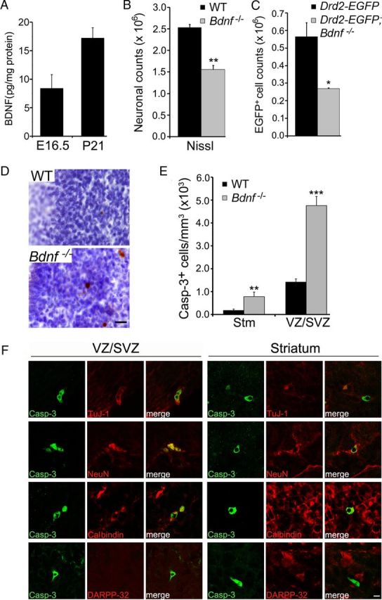
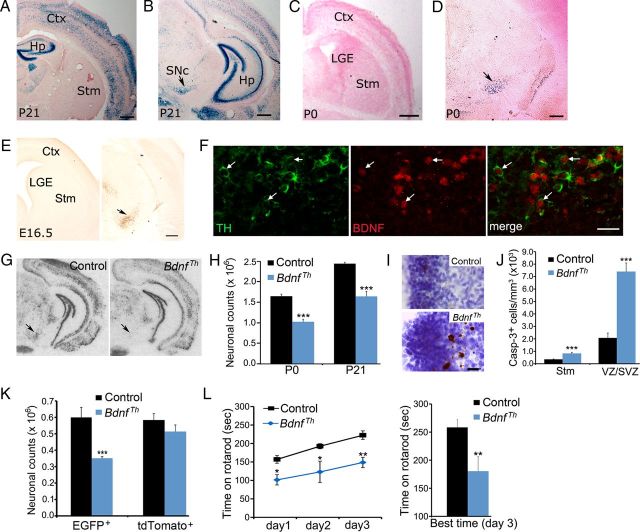
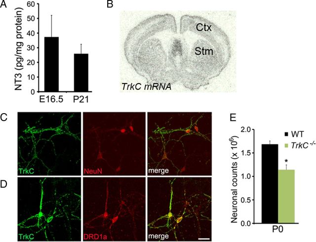
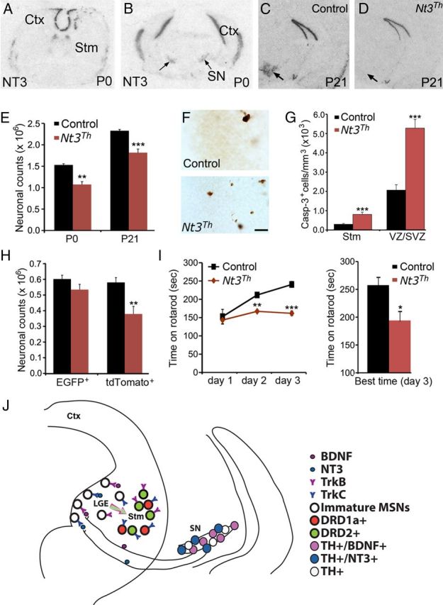
Neuronal death occurs at several stages during embryogenesis and early postnatal development; however, it is unknown how the survival of immature neurons at their origin is regulated before these cells migrate to their final destination. Striatal projection neurons, known as medium-sized spiny neurons (MSNs), in both the direct and indirect pathways are generated in the lateral ganglionic eminence (LGE). Here we report that brain-derived neurotrophic factor and neurotrophin-3 are anterogradely transported from midbrain dopaminergic neurons and support the survival of immature MSNs of the indirect and direct pathways, respectively, in the developing mouse striatum and LGE. These results reveal a novel mode of neurotrophic action in the nervous system by linking neurotrophins to the survival of immature neurons at their origin, while also suggesting that innervating neurons may control the size of their targeting neuronal population in the brain.


Figures

Figure 1.
Figure 1.
BDNF promotes survival of immature MSNs of the indirect pathway. A, BDNF protein levels in the striatum at E16.5 and P21. B, Counts of striatal neurons in WT and Bdnf −/− mice at P10–P14 (n = 3 for each genotype). C, Counts of striatal DRD2-expressing cells in Drd2-EGFP and Drd2-EGFP;Bdnf−/− mice at P10–P14 (n = 3–4 per genotype). D, Immunohistochemistry of activated caspase-3, counter-stained with Nissl. Scale bar, 25 μm. E, Density of cells containing activated caspase-3 in the striatum and the LGE VZ/SVZ of WT and Bdnf −/− mice at P0 (n = 3). F, Colocalization of caspase-3-positive cells with specific neuronal markers in the LGE VZ/SVZ and the striatum of Bdnf −/− mice at P0. Scale bar, 5 μm. Error bars indicate SEs. *p < 0.05; **p < 0.01; ***p < 0.001.
Figure 2.
Figure 2.
Midbrain dopaminergic neurons are the source of BDNF required for survival of MSNs of the indirect pathway. A–D, BDNF expression in BdnfLacZ/+ brain at P0 and P21, as revealed with X-gal staining for β-galactosidase (blue). Scale bars: A–C, 500 μm; D, 250 μm. E, BDNF expression in BdnfLacZ/+ embryos at E16.5, as revealed with immunohistochemistry of β-galactosidase. The arrow denotes the substantia nigra. Scale bar, 250 μm. F, Colocalization of TH with β-galactosidase in the substantia nigra of BdnfLacZ/+ embryos at E16.5. Arrows denote representative neurons expressing both TH and BDNF. Scale bar, 25 μm. G, Th-Cre-mediated deletion of the Bdnf gene. Arrows denote the substantia nigra. H, Counts of Nissl-stained striatal neurons in control and BdnfTh mice at P0 and P21 (n = 3–5 mice per group). I, Immunohistochemistry of activated caspase-3 in control and BdnfTh mice at P0. Scale bar, 25 μm. J, Density of cells containing activated caspase-3 in the striatum and the LGE of control and BdnfTh mice at P0 (n = 3–5). K, Counts of EGFP- and tdTomato-expressing striatal neurons in control and BdnfTh mice harboring either Drd2-EGFP or Drd1a-tdTomato at P21 (n = 3–5 mice per group). L, Control and BdnfTh mice (n = 11 and 7, respectively) at 6 months of age were tested on a rotarod. Data represent averages of scores from three trials each day for three consecutive days (left) and best score on the third day (right). Ctx, Cerebral cortex; Hp, hippocampus; SNc, substantia nigra pars compacta; Stm, striatum. Error bars indicate SEs. *p < 0.05; **p < 0.01; ***p < 0.001.
Figure 3.
Figure 3.
NT3-to-TrkC signaling is important for survival of striatal neurons. A, NT3 protein levels in the striatum were measured at E16.5 and P21. B, Radioactive in situ hybridization shows TrkC mRNA expression in the adult striatum (Stm) and cortex (Ctx). C, Many cultured striatal neurons, labeled by an antibody to neuronal marker (NeuN), express TrkC at 14 d in vitro. D, The majority of the MSNs of the direct pathway, labeled by an antibody to DRD1a, express TrkC. Scale bar, 25 μm. E, Counts of striatal neurons from Nissl-stained sections in WT and TrkC−/− mice at P0 (n = 3). Error bars indicate SEs. *p < 0.05.
Figure 4.
Figure 4.
NT3 from midbrain dopaminergic neurons promotes survival of immature MSNs of the direct pathway. A, B, Distribution of Ntf3 mRNA in the WT brain at P0. Arrows denote the substantia nigra. C, D, In situ hybridization shows the absence of Ntf3 mRNA in the substantia nigra (arrows) of Nt3Th mice at P21. E, Counts of Nissl-stained striatal neurons in control and Nt3Th mice at P0 and P21 (n = 3–5 mice per group). F, Immunohistochemistry of activated caspase-3 revealed apoptotic cells in the LGE VZ/SVZ of control and Nt3Th mice at P0. Scale bar, 25 μm. G, Density of cells containing activated caspase-3 in the striatum and the LGE of control and Nt3Th mice at P0 (n = 3–4). H, Counts of EGFP- and tdTomato-expressing striatal cells in control and Nt3Th mice harboring either Drd2-EGFP or Drd1a-tdTomoato at P21 (n = 3–5). I, Nt3Th mice (n = 8) exhibited poor motor performance and impaired motor learning compared with control mice (n = 7), assessed by rotarod tests. J, A model showing that BDNF and NT3 anterogradely transported from mesencephalic dopaminergic neurons regulate survival of immature neurons in the indirect and direct pathways, respectively. Ctx, Cerebral cortex; Stm, striatum; SN, substantia nigra. Error bars indicate SEs. *p < 0.05; **p < 0.01; ***p < 0.001.

References

    1. Alcántara S, Frisén J, del Río JA, Soriano E, Barbacid M, Silos-Santiago I. TrkB signaling is required for postnatal survival of CNS neurons and protects hippocampal and motor neurons from axotomy-induced cell death. J Neurosci. 1997;17:3623–3633. - PMC - PubMed
    1. Altar CA, Cai N, Bliven T, Juhasz M, Conner JM, Acheson AL, Lindsay RM, Wiegand SJ. Anterograde transport of brain-derived neurotrophic factor and its role in the brain. Nature. 1997;389:856–860. - PubMed
    1. Baydyuk M, Russell T, Liao GY, Zang K, An JJ, Reichardt LF, Xu B. TrkB receptor controls striatal formation by regulating the number of newborn striatal neurons. Proc Natl Acad Sci U S A. 2011;108:1669–1674. - PMC - PubMed
    1. Bolam JP, Hanley JJ, Booth PA, Bevan MD. Synaptic organisation of the basal ganglia. J Anat. 2000;196:527–542. - PMC - PubMed
    1. Danielian PS, Muccino D, Rowitch DH, Michael SK, McMahon AP. Modification of gene activity in mouse embryos in utero by a tamoxifen-inducible form of Cre recombinase. Curr Biol. 1998;8:1323–1326. - PubMed

Publication types

MeSH terms

LinkOut - more resources