. 2013 Feb 20;33(8):3646-58.
doi: 10.1523/JNEUROSCI.1635-12.2013.

CCAAT enhancer binding protein δ plays an essential role in memory consolidation and reconsolidation

Affiliations

CCAAT enhancer binding protein δ plays an essential role in memory consolidation and reconsolidation

Amy A Arguello et al. J Neurosci. .

Abstract

A newly formed memory is temporarily fragile and becomes stable through a process known as consolidation. Stable memories may again become fragile if retrieved or reactivated, and undergo a process of reconsolidation to persist and strengthen. Both consolidation and reconsolidation require an initial phase of transcription and translation that lasts for several hours. The identification of the critical players of this gene expression is key for understanding long-term memory formation and persistence. In rats, the consolidation of inhibitory avoidance (IA) memory requires gene expression in both the hippocampus and amygdala, two brain regions that process contextual/spatial and emotional information, respectively; IA reconsolidation requires de novo gene expression in the amygdala. Here we report that, after IA learning, the levels of the transcription factor CCAAT enhancer binding protein δ (C/EBPδ) are significantly increased in both the hippocampus and amygdala. These increases are essential for long-term memory consolidation, as their blockade via antisense oligodeoxynucleotide-mediated knockdown leads to memory impairment. Furthermore, C/EBPδ is upregulated and required in the amygdala for IA memory reconsolidation. C/EBPδ is found in nuclear, somatic, and dendritic compartments, and a dendritic localization of C/EBPδ mRNA in hippocampal neuronal cultures suggests that this transcription factor may be translated at synapses. Finally, the induction of long-term potentiation at CA3-CA1 synapses by tetanic stimuli in acute slices, a cellular model of long-term memory, leads to an accumulation of C/EBPδ in the nucleus. We conclude that the transcription factor C/EBPδ plays a critical role in memory consolidation and reconsolidation.


Figures
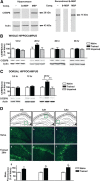
Figure 1.
Figure 1.
Hippocampal C/EBPδ level is increased after IA training. A, Validation of C/EBPδ antibody specificity for Western blotting. Western blot immunostaining of hippocampal extracts (H) or recombinant CEBPδ-MBP (δ-MBP, R) incubated with C/EBPδ antibody in the presence of 200× excess of either δ-MBP or MBP. Both the 29 kDa band in H and the 75 kDa δ-MBP in R were competed by δ-MBP but not MBP. B, Densitometric analysis of C/EBPδ in whole hippocampal extracts via quantitative Western blotting, with representative blots shown below each graph. Rats were trained and killed at different time points after training. C/EBPδ levels remain unchanged at 12, 36, or 96 h after training compared with naive or unpaired controls. However, a trend toward an increase was seen at 20 h after training (naive, n = 3 or 4; unpaired, n = 4 or 5; trained, n = 4 or 5 for all time points). Data are expressed as mean percentage ± SEM of naive (100%) control mean values. C, Densitometric analysis of C/EBPδ in dorsal hippocampal extracts with representative blots shown below each graph. C/EBPδ levels were significantly increased at 20 h after training compared with naive and unpaired controls. *p < 0.05. **p < 0.01. No change was found at 0.5 or 12 h after training (naive, n = 5 or 6; unpaired, n = 4–6; trained, n = 4–6 for all time points). Data are expressed as mean percentage ± SEM of naive (100%) control mean values. D, Immunofluorescence of C/EBPδ staining. Schematic representations of the areas shown in the pictures are indicated in the green boxes. Examples of DG, CA1, and CA3 of naive (a–c) and trained rats (d–f) showing an induction of C/EBPδ 20 h after training (n = 4/group). g–i, Quantitative mean fluorescence analysis revealed a significant increase in C/EBPδ in all subregions. Scale bar, 100 μm.
Figure 2.
Figure 2.
Hippocampal C/EBPδ protein is required for the consolidation of IA memory. A, Rats were injected with SC-ODN or δ-ODN 5 h after training and tested 48 h after training. Memory retention was comparable in both groups (n = 7 or 8). Retentions are expressed as mean latency ± SEM in seconds. B, Rats injected with δ-ODN 12 h after training showed a significant memory impairment compared with SC-ODN-injected rats at 48 h after training (n = 8/group). *p < 0.05. Retentions are expressed as mean latency ± SEM in seconds. C, Rats injected with δ-ODN 12 h after training showed no difference in locomotor activity and anxiety-like activity, measured by entries into and time spent in the center zone, compared with SC-ODN-injected rats in the open field test at 48 h after training (n = 8 or 9/group).
Figure 3.
Figure 3.
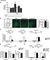
C/EBPδ in the BLA is upregulated after training and required for the consolidation and reconsolidation of IA memory. A, Quantitative Western blot analysis of C/EBPδ in BLA extracts with representative blots shown below each graph. C/EBPδ levels were significantly increased at 9 and 20 h after training compared with naive controls: *p < 0.05; ***p < 0.001. No significant change was found at 0.5, 6, or 48 h after training (naive, n = 22; 0.5 h, n = 10; 6 h, n = 7; 9 h, n = 7; 20 h, n = 13; 48 h, n = 11). C/EBPδ level at 9 h after unpaired protocol was not different from that of naive conditions but significantly different from the trained (n = 6). Data are expressed as mean percentage ± SEM of naive (100%) control mean values. B, Representative examples of C/EBPδ immunohistochemical staining in the BLA of naive, unpaired, or IA trained rats. Scale bar, 200 μm. Quantitative analyses of C/EBPδ fluorescence intensity showed a significant increase of C/EBPδ levels in the BLA of rats 20 h after training compared with naive and unpaired controls killed at the matched time point (n = 4/group): ***p < 0.001. C, Rats were injected with SC-ODN or δ-ODN 5 or 12 h after training and were tested 48 h after training. In both cases, δ-ODN profoundly disrupted memory retention compared with SC-ODN (SC-ODN, n = 7–9; δ-ODN, n = 6 or 7): ***p < 0.0001; *p < 0.05. In contrast, SC-ODN or δ-ODN injections 48 h after training did not affect memory tested 96 h after training (SC-ODN, n = 9; δ-ODN, n = 10). Retentions are expressed as mean latency ± SEM in seconds. D, Rats injected with δ-ODN 12 h after training showed no difference in locomotor activity and anxiety-like activity, measured by entries into and time spent in the center zone, compared with SC-ODN-injected rats in the open field test at 48 h after training (n = 8–10/group). E, Rats injected with δ-ODN 5 h after memory reactivation (Test 1, R, δ-ODN, n = 8) showed significantly lower latencies when retested 48 h later, compared with rats injected with SC-ODN (R, SC-ODN, n = 8) or rats injected with δ-ODN at the matching time point in the absence of memory reactivation (NR, δ-ODN, n = 7).
Figure 4.
Figure 4.
C/EBPδ immunofluorescent staining. A, Representative immunofluorescence of C/EBPδ (green) and the dendritic marker MAP2 (red) on brain sections taken 20 h after training. a–c, Hippocampus. e–g, Amygdala, i–k, Cerebellum. m–o, Cortex. Scale bar, 50 μm. c, g, k, o, Boxed regions are shown at high magnification of d, h, l, and p, respectively. Scale bar, 15 μm. B, Representative immunofluorescence of C/EBPδ and MAP2 on hippocampal neuronal cultures, and validation of C/EBPδ antibody specificity for immunofluorescent staining. a–c, −, no competition. C/EBPδ immunofluorescence (green) detected by anti-C/EBPδ is evident in the somatic and dendritic (MAP2-positive, red) compartments. d–f, Twenty × excess of δ-MBP incubated with anti-C/EBPδ competed for C/EBPδ (green) but not for MAP2 (red) immunofluorescence. g–i, Twenty times excess of MBP (control) did not compete for C/EBPδ (green) or MAP2 (red) immunofluorescence. Scale bar, 25 μm.
Figure 5.
Figure 5.
C/EBPδ FISH. A, Hippocampal CA1 (a–d) and insular cortical sections (e–h) taken 20 h after training were hybridized with control C/EBPδ sense probe (a, e) or C/EBPδ antisense probe (b, f). Dendritic regions of brain sections shown in b and f are revealed by MAP2 immunostaining (c, g). Merged images of C/EBPδ mRNA (green) and MAP2 (red) show primary somatic localization of C/EBPδ mRNA in adult rat brain sections. Scale bar, 25 μm. B, C/EBPδ FISH of hippocampal neuronal cultures. a, Control C/EBPδ sense probe. b, Antisense probe to C/EBPδ. c, MAP2 immunofluorescence. d, Merged image of C/EBPδ mRNA (green) and MAP2 (red) reveals the localization of C/EBPδ mRNA in both somatic and dendritic compartments of cultured hippocampal neurons. Scale bar, 12.5 μm.
Figure 6.
Figure 6.
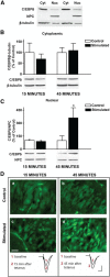
C/EBPδ expression is induced after LTP. A, Representative Western blots validating cytoplasmic (Cyt) and nuclear (Nuc) fractionation of C/EBPδ expression levels. NPC, immunostaining with anti-NPC is enriched in the nuclear fraction; β-tubulin, immunostaining with anti-β-tubulin is enriched in the cytoplasmic fraction. B, Quantitative Western blot analyses of cytoplasmic fractions from hippocampal slices processed 15 min or 45 after LTP induction; C/EBPδ protein levels remain unchanged. Representative Western blot examples are shown below each graph. Data are expressed as mean percentage ± SEM of unstimulated slice extract (control) mean values (n = 4/group). C, Quantitative Western blot analyses of nuclear fractions from hippocampal slices processed 15 or 45 min after LTP induction; C/EBPδ levels significantly increase in the nuclear fraction at 45 min but not at 15 min after LTP induction. Representative Western blot examples are shown below each graph. Data are expressed as mean percentage ± SEM of unstimulated slice extract (control) mean values (n = 4/group). D, Examples of anti-C/EBPδ immunofluorescence staining showing no change in C/EBPδ levels 15 min after LTP induction (c) compared with unstimulated control (a). C/EBPδ immunofluorescence increases 45 min after LTP induction (d) compared with unstimulated control (b) (n = 4/group). Scale bar, 20 μm. Bottom, Representative traces from LTP-elicited slices. Traces were recorded before and 15 (c) or 45 min (d) after LTP induction. Calibration: 10 ms, 0.5 mV.

References

    1. Alberini CM. The role of protein synthesis during the labile phases of memory: revisiting the skepticism. Neurobiol Learn Mem. 2008;89:234–246. - PMC - PubMed
    1. Alberini CM. Transcription factors in long-term memory and synaptic plasticity. Physiol Rev. 2009;89:121–145. - PMC - PubMed
    1. Alberini CM. The role of reconsolidation and the dynamic process of long-term memory formation and storage. Front Behav Neurosci. 2011;5:12. - PMC - PubMed
    1. Alberini CM, Ghirardi M, Metz R, Kandel ER. C/EBP is an immediate-early gene required for the consolidation of long-term facilitation in Aplysia. Cell. 1994;76:1099–1114. - PubMed
    1. Alberini CM, Ghirardi M, Huang YY, Nguyen PV, Kandel ER. A molecular switch for the consolidation of long-term memory: cAMP-inducible gene expression. Ann N Y Acad Sci. 1995;758:261–286. - PubMed

Publication types

Substances

LinkOut - more resources