. 2013 Jul;28(7):1631-40.
doi: 10.1002/jbmr.1894.

Periapical disease and bisphosphonates induce osteonecrosis of the jaws in mice

Affiliations

Periapical disease and bisphosphonates induce osteonecrosis of the jaws in mice

Ben Kang et al. J Bone Miner Res. 2013 Jul.

Abstract

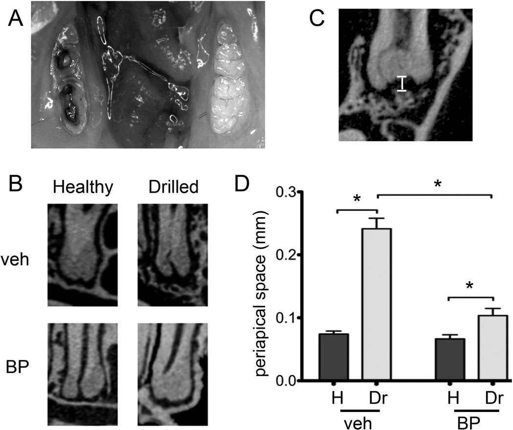
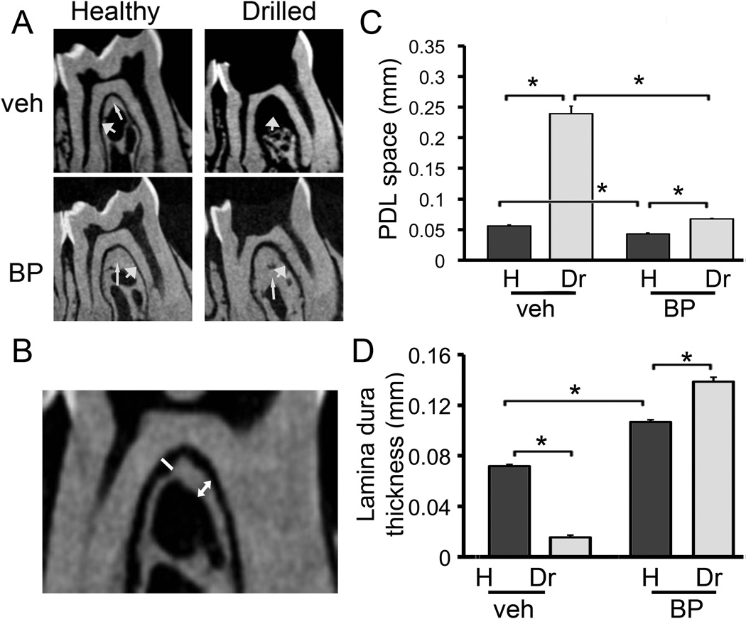
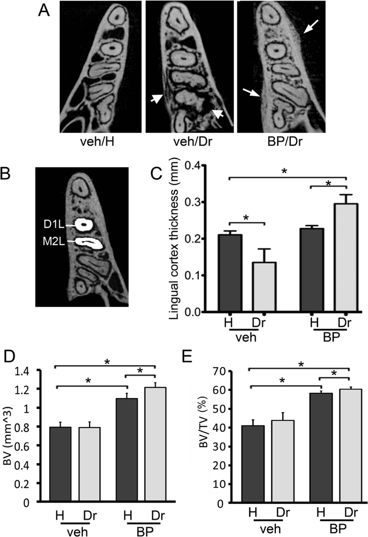
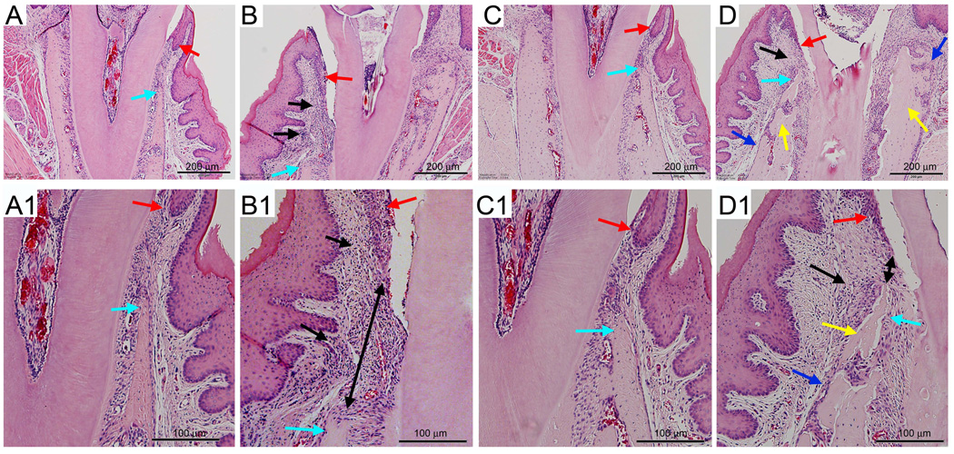
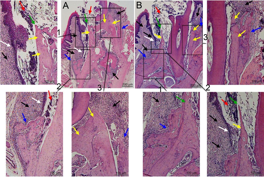
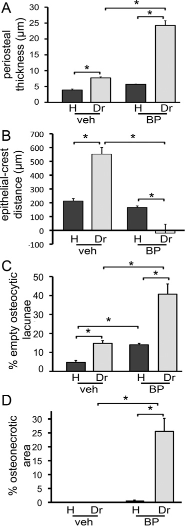
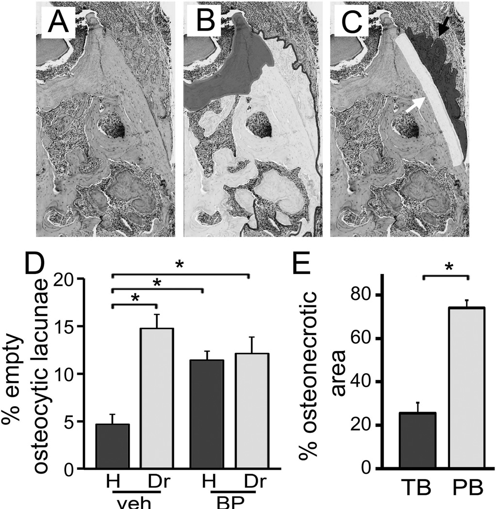
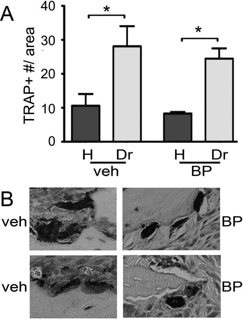
Osteonecrosis of the jaw (ONJ) is a well-recognized complication of antiresorptive medications, such as bisphosphonates (BPs). Although ONJ is most common after tooth extractions in patients receiving high-dose BPs, many patients do not experience oral trauma. Animal models using tooth extractions and high BP doses recapitulate several clinical, radiographic, and histologic findings of ONJ. We and others have reported on rat models of ONJ using experimental dental disease in the absence of tooth extraction. These models emphasize the importance of dental infection/inflammation for ONJ development. Here, we extend our original report in the rat, and present a mouse model of ONJ in the presence of dental disease. Mice were injected with high dose zoledronic acid and pulpal exposure of mandibular molars was performed to induce periapical disease. After 8 weeks, quantitative and qualitative radiographic and histologic analyses of mouse mandibles were done. Periapical lesions were larger in vehicle-treated versus BP-treated mice. Importantly, radiographic features resembling clinical ONJ, including thickening of the lamina dura, periosteal bone deposition, and increased trabecular density, were seen in the drilled site of BP-treated animals. Histologically, osteonecrosis, periosteal thickening, periosteal bone apposition, epithelial migration, and bone exposure were present in the BP-treated animals in the presence of periapical disease. No difference in tartrate-resistant acid phosphatase (TRAP)+ cell numbers was observed, but round, detached, and removed from the bone surface cells were present in BP-treated animals. Although 88% of the BP-treated animals showed areas of osteonecrosis in the dental disease site, only 33% developed bone exposure, suggesting that osteonecrosis precedes bone exposure. Our data further emphasize the importance of dental disease in ONJ development, provide qualitative and quantitative measures of ONJ, and present a novel mouse ONJ model in the absence of tooth extraction that should be useful in further exploring ONJ pathophysiological mechanisms.


Conflict of interest statement

DISCLOSURES

Dr. Tetradis has served as a paid consultant for and has received grant support from Amgen Inc. All other authors state that they do not have any conflicts of interest.

Figures

Figure 1
Figure 1
Experimental periapical disease model. A) The crowns of the first and second molars were drilled to create pulp exposure and cause periapical disease in veh or BP treated animals. B) Representative µCT images of the periapical area of the first molar distal root of healthy and drilled sites in veh or BP treated animals are shown. C) To quantify periapical bone loss, the periapical space was measured as the distance from the root apex to the periapical alveolar bone. D) Periapical space at the distal root of the first molar (D1). * statistically significantly different, p<0.01.
Figure 2
Figure 2
Changes in the furcational periodontium of veh and BP treated animals. A) Representative µCT images of the furcation area of the first molar of healthy and drilled sites in veh or BP treated animals are shown. Thin arrow points to the lamina dura and thick arrow points to the PDL space. B) To quantify changes in the furcational periodontium, the width of the PDL space and the thickness of the lamina dura were measured at the furcation area. C) PDL space at the furcation area of the first molar. * statistically significantly different, p<0.01. D) Lamina dura thickness at the furcation area of the first molar. * statistically significantly different, p<0.01.
Figure 3
Figure 3
Changes in alveolar bone structure in veh and BP treated animals. A) Representative axial µCT slices of the alveolar ridge at the apical third of the root are shown. Thick arrows point to osteolysis extending to the mandibular cortices seen in the drilled site of the veh treated animals. Thin arrows point to periosteal bone deposition and increased trabecular density seen in BP treated animals. B) To quantify the lingual bone thickness in veh vs. BP treated animals, the lingual width of the alveolar bone was measured at the distal root of the first moral and mesial root of the second molar in the healthy and drilled sites. C) Lingual cortex thickness at the distal root of the first molar. D) BV and E) BV/TV of bone at the area of the alveolar ridge. * statistically significantly different, p<0.05.
Figure 4
Figure 4
Histologic examination of periodontal area of the alveolar bone. A, A1) Healthy site of a veh treated animal. B, B1) Drilled site of a veh treated animal. C, C1) Healthy site of a BP treated animal. D, D1) Drilled site of a BP treated animal. A, B, C, D are at 4X magnification, while A1, B1, C1, and D1 demonstrate a magnified area of A, B, C, and D. Red arrows point to marginal gingival epithelium, aqua to the crestal alveolar bone, black arrows to inflammatory infiltrate, black double arrows to the epithelial-crestal bone distance, yellow arrows to necrotic bone, and blue arrows to periosteal bone deposition.
Figure 5
Figure 5
Histologic examination of the drilled site of two BP treated animals (A and B) with bone exposure. A and B are at 4X magnification, while insets demonstrate magnified areas of A and B. Black arrows point to inflammatory infiltrate, white arrows to epithelial migration, yellow arrows to necrotic bone, green arrows to exposed bone, red arrows to debris, and blue arrows to periosteal bone deposition.
Figure 6
Figure 6
Quantification of histologic findings. A) Thickness of periosteum at the buccal and lingual alveolus was measured. B) The shortest epithelial-crest distance was determined. If epithelium extended below the level of the alveolar crest, a negative value was assigned to the measurement. C) Osteocytic lacunae were measured and empty lacunae were expressed as percent of total. D) Area of osteonecrosis was measured and expressed as % of total bone area. * statistically significantly different, p<0.01.
Figure 7
Figure 7
Quantification of BP effects on empty osteocytic lacunae and osteonecrosis. A) Baseline histologic appearance of the area of interest. B) The necrotic (artificially shaded dark grey) and viable (artificially shaded white) bone areas were identified. C) The periosteal bone deposition (artificially shaded dark grey and black arrow) and adjacent 50 µm wide (artificially shaded white and white arrow) bone area were established. D) Osteocytic lacunae were measured in the areas of viable bone and empty lacunae were expressed as percent of total. E) The total bone % osteonecrotic area (TB) and % osteonecrotic area in a 50 µm wide band adjacent to periosteal bone (PB) were measured. * statistically significantly different, p<0.01.
Figure 8
Figure 8
Effects of BP treatment on TRAP+ cells. A) Number of TRAP+ cells in the healthy and drilled site of veh and BP treated animals. * statistically significantly different, p<0.01. B) Representative examples of TRAP+ cells from the drilled site of veh or BP treated animals.

References

    1. Allen MR. Bisphosphonates and osteonecrosis of the jaw: moving from the bedside to the bench. Cells Tissues Organs. 2009;189:289–294. - PMC - PubMed
    1. Russell RG. Bisphosphonates: the first 40 years. Bone. 2011;49:2–19. - PubMed
    1. Russell RG, Watts NB, Ebetino FH, Rogers MJ. Mechanisms of action of bisphosphonates: similarities and differences and their potential influence on clinical efficacy. Osteoporos Int. 2008;19:733–759. - PubMed
    1. Roelofs AJ, Thompson K, Gordon S, Rogers MJ. Molecular mechanisms of action of bisphosphonates: current status. Clin Cancer Res. 2006;12:6222s–6230s. - PubMed
    1. Russell RG, Rogers MJ. Bisphosphonates: from the laboratory to the clinic and back again. Bone. 1999;25:97–106. - PubMed

Publication types

MeSH terms