. 2013 Jul;28(7):1587-98.
doi: 10.1002/jbmr.1901.

In vivo overexpression of tissue-nonspecific alkaline phosphatase increases skeletal mineralization and affects the phosphorylation status of osteopontin

Affiliations

In vivo overexpression of tissue-nonspecific alkaline phosphatase increases skeletal mineralization and affects the phosphorylation status of osteopontin

Sonoko Narisawa et al. J Bone Miner Res. 2013 Jul.

Abstract

Functional ablation of tissue-nonspecific alkaline phosphatase (TNAP) (Alpl⁻/⁻ mice) leads to hypophosphatasia, characterized by rickets/osteomalacia attributable to elevated levels of extracellular inorganic pyrophosphate, a potent mineralization inhibitor. Osteopontin (OPN) is also elevated in the plasma and skeleton of Alpl⁻/⁻ mice. Phosphorylated OPN is known to inhibit mineralization, however, the phosphorylation status of the increased OPN found in Alpl⁻/⁻ mice is unknown. Here, we generated a transgenic mouse line expressing human TNAP under control of an osteoblast-specific Col1a1 promoter (Col1a1-Tnap). The transgene is expressed in osteoblasts, periosteum, and cortical bones, and plasma levels of TNAP in mice expressing Col1a1-Tnap are 10 to 20 times higher than those of wild-type mice. The Col1a1-Tnap animals are healthy and exhibit increased bone mineralization by micro-computed tomography (µCT) analysis. Crossbreeding of Col1a1-Tnap transgenic mice to Alpl⁻/⁻ mice rescues the lethal hypophosphatasia phenotype characteristic of this disease model. Osteoblasts from [Col1a1-Tnap] mice mineralize better than nontransgenic controls and osteoblasts from [Col1a1-Tnap⁺/⁻; Alpl⁻/⁻] mice are able to mineralize to the level of Alpl⁺/⁻ heterozygous osteoblasts, whereas Alpl⁻/⁻ osteoblasts show no mineralization. We found that the increased levels of OPN in bone tissue of Alpl⁻/⁻ mice are comprised of phosphorylated forms of OPN whereas wild-type (WT) and [Col1a1-Tnap⁺/⁻; Alpl⁻/⁻] mice had both phosphorylated and dephosphorylated forms of OPN. OPN from [Col1a1-Tnap] osteoblasts were more dephosphorylated than nontransgenic control cells. Titanium dioxide-liquid chromatography and tandem mass spectrometry analysis revealed that OPN peptides derived from Alpl⁻/⁻ bone and osteoblasts yielded a higher proportion of phosphorylated peptides than samples from WT mice, and at least two phosphopeptides, p(S¹⁷⁴FQVS¹⁷⁸DEQY¹⁸²PDAT¹⁸⁶DEDLT¹⁹¹)SHMK and FRIp(S²⁹⁹HELES³⁰⁴S³⁰⁵S³⁰⁶S³⁰⁷)EVN, with one nonlocalized site each, appear to be preferred sites of TNAP action on OPN. Our data suggest that the promineralization role of TNAP may be related not only to its accepted pyrophosphatase activity but also to its ability to modify the phosphorylation status of OPN.


Conflict of interest statement

Conflict of interest: All authors report no conflicts of interest.

Figures

Figure 1
Figure 1. Construction and characterization of Col1a1-Tnap mice
(A) The 5.3 kb transgene fragment consists of a 2.3 kb Col1a1 promoter, an intron, a full TNAP cDNA (2.5 kb) and a polyA sequence isolated from the pStec1 backbone vector by Sac I digestion.(36) The 2.3 kb Col1a1 promoter was isolated from the pJ251 vector (a kind gift from Dr. Gerard Karsenty, Columbia University, NY) and the 2.5 kb fragment of human ALPL cDNA was derived from pSV2Aalp (ATCC 59634). The purified DNA construct was used for microinjection into the pronucleus of embryos from FVB/N mice in the transgenic facility at the Sanford-Burnham Medical Research Institute. (B) Integration of transgene was analyzed by Southern hybridization using standard protocols. Four micrograms of tail DNA was digested with Hind III and probed with 675 bp fragment from intron 2 of Alpl gene together with a 498 bp Bgl I fragment from the human ALPL cDNA. The transgene was transmitted to the offspring in a Mendelian pattern. (C) Body weight at four months of age: female Col1a1-Tnap+/− or Col1a1-Tnap+/+ (ColTg) vs WT p=0.5401, female WT vs [Col1a1-Tnap+/−; Alpl−/−] (ColTg; Alpl−/−) p=0.0276. The body weight difference is more pronounced in female than male animals. (D) An average TNAP value from triplicated measurement for each animal was plotted in the Fig. D, (numbers of animals, Col1a1-Tnap+/−, Col1a1-Tnap+/+, WT and Col1a1-Tnap−/−, are n=18, 7, 11, 5 respectively). Plasma AP levels display a wide range but are consistently elevated in the transgenic mice. Col1a1-Tnap+/− or Col1a1-Tnap+/+: 504 ± 261 μg/mL, WT: 25.8 ± 14.7 μg/mL, [Col1a1-Tnap+/− or Col1a1-Tnap+/+; Alpl−/−]: 478 ± 221 μg/mL. The difference between Col1a1-Tnap+/− (hemizygous) and Col1a1-Tnap+/+ (homozygous) was not significant (p=0.3967), while hemizygous Tg display a significant increase in AP compared to WT (p<0.0001).
Figure 2
Figure 2. AP activity staining on tissues from 11-day-old mice
A, B, C and D: wild type. E, F, G and H: [ApoE-Tnap+/−; Alpl−/−]. I, J, K and L: [Col1a1-Tnap+/−; Alpl−/−]. M, N, O and P: Alpl−/− mice. A, E, I and M: fibula. B, E, H and K: calvaria and forebrain. C, G, K and O: liver. D, H, L and P: kidney and adrenal gland. Organs for each genotype shown are derived from a single animal. Two to four mice per each genotype were analyzed and typical results were shown. (×100)
Figure 3
Figure 3. PPi and OPN measurement
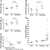
A and C: plasma PPi. B and D: plasma OPN. A and B: four-month-old Col1a1-Tnap+/−, WT and [Col1a1-Tnap+/−; Alpl−/−] mice. C: four-month-old ApoE-Tnap+/−, WT and [ApoE-Tnap+/−; Alpl−/−] mice. D: 11-day-old Col1a1-Tnap+/−, WT, [Col1a1-Tnap+/−; Alpl−/−] and Alpl−/− mice. E: correlation between body weight and plasma OPN in 11-day-old Alpl−/− mice. An average value from duplicated assay for each mouse was plotted in the figures. (A, B and C: n=5, D: n=4 except that Alpl−/− was n=18).
Figure 4
Figure 4. Mineralization assay
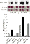
Osteoblasts were isolated from calvarial bones of two-day-old mice as described.(8) Culture media containing 100 μg/mL ascorbic acid and 2 or 10 mM ®GP was renewed every second day and the cells were fixed in 100% ethanol 19 days later for AP staining and mineralization assay.(41) A western blot to detect TNAP is shown in the top row. The sample from [Col1a1-Tnap+/−; Alpl−/−] (ColTg; Alpl-/-) represents human TNAP protein expressed from the transgene and the sample from Alpl+/− control shows endogenous mouse TNAP. Human and mouse TNAP share 91.5 % homology in their peptide sequences, and the rat monoclonal antibody used in this study recognizes both human and mouse TNAP. Cells of each genotype were derived from a single newborn pup. The same experiment was repeated twice to test reproducibility.
Figure 5
Figure 5. Bone histomorphometric analysis
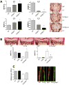
A: comparison of area of mineralized bone (BV/TV) and unmineralized osteoind (OV/BV) in L2 and L3 vertebrae bones from four-month-old WT, Col1a1-Tnap+/−, and Col1a1-Tnap+/+ mice (n=3). B: comparison of area of mineralized bone (BV/TV) and unmineralized osteoind (OV/BV) in tibia from 16-day-old WT, Col1a1-Tnap+/−, Alpl−/− and [Col1a1-Tnap+/−; Alpl−/−] mice (n=3). C: mineral apposition rates in parietal bones from 11-day-old WT, Col1a1-Tnap+/−, Alpl−/− and [Col1a1-Tnap+/−; Alpl−/−] mice (n=3)
Figure 6
Figure 6. Western blotting
Thirty nanograms of recombinant mouse OPN (Sigma, Saint Louis, MO, USA) was digested with 10 U of IAP in AP reaction buffer for two hours and used as a control of dephosphorylated OPN (r.mOPN+IAP). A: cultured osteoblasts under mineralizing condition. B: bone tissue. The same membranes were re-probed for β-actin. C: Immunoprecipitation experiment. Untreated and IAP treated samples were compared by western blot with goat anti-OPN antibody (Figure 6C left and right). D: Comparison of OPN migration in Phos-Tag SDS PAGE. Left: osteoblast extracts. Right: bone extracts from 11-day-old and four-month-old. Western blots with anti-OPN antibody were shown along with Ponceau S staining of the same membrane to indicate the total protein blotted. Samples in each lane were derived from a single mouse and these experiments were repeated at least 2 times with samples derived from different animals.
Figure 7
Figure 7. Bone phenotype of [Col1a1-Tnap+/−; Alpl−/−]
A: X-ray image of hind limbs. [Col1a1-Tnap+/−; Alpl−/−] (left) and WT (right). B: Higher magnification of X-ray images of knee joints. Top [Col1a1-Tnap+/−; Alpl+/+], middle WT and bottom [Col1a1-Tnap+/−; Alpl−/−]. C, D and E are von Kossa staining on undecalcified femur sections from [Col1a1-Tnap+/−; Alpl+/+], WT and [Col1a1-Tnap+/−; Alpl−/−], respectively. F, G and H: WT control. I, J and K: [Col1a1-Tnap+/−; Alpl−/−]. F and I: distal femur (×40). G, H, J and K: proximal tibia (×400). F, G, I and J: Goldner's trichrome staining. H and K: immunohistochemistry showing expression of endogenous mouse TNAP in WT control (H) and human TNAP in [Col1a1-Tnap+/−; Alpl−/−] (K). G H, and J K are serial sections respectively. The epiphyseal plate is almost missing from some areas of the tibia and femur of [Col1a1-Tnap+/−; Alpl−/−] mice (Figure 7I). The proliferating chondrocyte zone has an abnormal structure (arrows in Figure 7J) and formation of trabecular bones is also reduced in the adult [Col1a1-Tnap+/−; Alpl−/−] mice (Figure 7J). Endogenous TNAP is highly expressed near the hypertrophic zone (Figure 7H), while the expression of transgenic TNAP is limited (Figure 7K).

References

    1. Narisawa S, Hasegawa H, Watanabe K, Millán JL. Stage-specific expression of alkaline phosphatase during neural development in the mouse. Dev Dyn. 1994;201:227–35. - PubMed
    1. Narisawa S, Fröhlander N, Millán JL. Inactivation of two mouse alkaline phosphatase genes and establishment of a model of infantile hypophosphatasia. Dev Dyn. 1997;208:432–46. - PubMed
    1. Narisawa S, Wennberg S, Millán JL. Abnormal vitamin B6 metabolism in alkaline phosphatase knock-out mice causes multiple abnormalities, but not the impaired bone mineralization. J Pathol. 2001;193:125–33. - PubMed
    1. Fedde K, Blair L, Silverstein J, Coburn SP, Ryan LM, Weinstein RS, Waymire K, Narisawa S, Millán JL, MacGregor GR, Whyte MP. Alkaline phosphatase knock-out mice recapitulate the metabolic and skeletal defects of infantile hypophosphatasia. J Bone Miner Res. 1999;14:2015–25. - PMC - PubMed
    1. Anderson HC, Sipe JB, Hessle L, Dhamyamraju R, Atti E, Camacho NP, Millán JL. Impaired calcification around matrix vesicles of growth plate and bone in alkaline phosphatase- deficient mice. Am J Pathol. 2004;164:841–7. - PMC - PubMed

Publication types