. 2013 Jun 15;22(12):2376-86.
doi: 10.1093/hmg/ddt082. Epub 2013 Feb 19.

Tardbpl splicing rescues motor neuron and axonal development in a mutant tardbp zebrafish

Affiliations

Tardbpl splicing rescues motor neuron and axonal development in a mutant tardbp zebrafish

Channa A A Hewamadduma et al. Hum Mol Genet. .

Abstract

Mutations in the transactive response DNA binding protein-43 (TARDBP/TDP-43) gene, which regulates transcription and splicing, causes a familial form of amyotrophic lateral sclerosis (ALS). Here, we characterize and report the first tardbp mutation in zebrafish, which introduces a premature stop codon (Y220X), eliminating expression of the Tardbp protein. Another TARDBP ortholog, tardbpl, in zebrafish is shown to encode a Tardbp-like protein which is truncated compared with Tardbp itself and lacks part of the C-terminal glycine-rich domain (GRD). Here, we show that tardbp mutation leads to the generation of a novel tardbpl splice form (tardbpl-FL) capable of making a full-length Tardbp protein (Tardbpl-FL), which compensates for the loss of Tardbp. This finding provides a novel in vivo model to study TDP-43-mediated splicing regulation. Additionally, we show that elimination of both zebrafish TARDBP orthologs results in a severe motor phenotype with shortened motor axons, locomotion defects and death at around 10 days post fertilization. The Tardbp/Tardbpl knockout model generated in this study provides an excellent in vivo system to study the role of the functional loss of Tardbp and its involvement in ALS pathogenesis.


Figures

Figure 1.
Figure 1.
The tardbp fh301 mutant (tardbpfh301/+) zebrafish generated by a TILLING process is viable. (A) Through mutation screening of a zebrafish ENU mutagenesis library, a c.660 C>A missense in-frame mutation in tardbp was detected. (B) The c.660 C>A mutation results in a premature truncation mutation at the 220 amino acids residue (Threonine) in the RRM2 domain. (C) c.660 C>A also results in a loss of CViQ1 restriction digest enzyme site, which helps to identify the genotype of the zebrafish following fin clipping. (D) Diagrammatic representation of the measurement of SL of zebrafish at 6 months of age. (E) Fry from a tardbpfh301/+heterozygous in cross at F5 generation were fin clipped at 6 months of age to identify the genotype. The weights were significantly different between tardbp+/+ and tardbpfh301/fh301 (230 mg, SD ± 39 versus 183 mg, SD ± 40, P < 0.0001) and tardbpfh301/+ (215 mg, SD ± 46, P < 0.001). There was no significant weight difference between tardbpfh301/+and tardbp+/+ (P> 0.05). (F) Measurement of the SL revealed significant differences between tardbp+/+ (23.81 mm, SD ± 1.76), tardbpfh301/+ (22.83 mm, SD ± 1.26) and tardbpfh301/fh301 (21.67 mm, SD ± 1.84)(*P < 0.01, **P < 0.001, ***P < 0.0001). (G) An immunoblot probed with h.TDP-43 Ab2 (which binds to the C-terminus of TDP-43) demonstrates a loss of Tardbp from all tissues in a 6-month-old adult homozygous mutant zebrafish (tardbpfh301/fh301). h.TDP-43 Ab2 is specific to Tardbp and does not detect Tardbpl.
Figure 2.
Figure 2.
The tardbpfh301/fh301 null phenotype is rescued by over-expression of a tardbpl full-length protein (Tardbpl-FL). (A) Western blot of tissues from 6-month-old adult tardbpfh301/fh301 zebrafish using h.TDP-43 Ab1 which binds to the N-terminus of TDP-43. Compared with the tardbp+/+, tardbpfh301/fh301 embryos have an over expressed signal at ∼43 kDa molecular weight indicated by $. This relative over-expression of a protein similar to the molecular weight of Tardbp is present in all the tested tissues of tardbpfh301/fh301 adult zebrafish. (B) tardbpl AMO injection into tardbpfh301/fh301 fish resulted in near complete (5 ng) and complete (16 ng) knockdown of Tardbpl-FL over expression, and Tardbpl (∼33 kDa, indicated by ¥) suggesting that tardbpl and tardbpl-FL share a similar translational initiating region. (C) Tardbpl-FL protein is upregulated 20-fold in the tardbpfh301/fh301compared with the tardbp+/+(P < 0.001). (D) Tardbpl-FL and Tardbpl are knocked down by tardbpl AMO injection in tardbpfh301/fh301 embryos (P < 0.0001).
Figure 3.
Figure 3.
Effects of Tardbpl-FL knockdown in WT and tardbp null zebrafish embryos. (AD) Uninjected, control AMO (CoMo) injected and tardbpl AMO with p53 (tardbpl AMO +p53) groups of WT (tardbp+/+) embryos were morphologically normal when compared with tardbpl AMO with p53 injected HOM (tardbpfh301 /fh301 embryos) zebrafish (EH) which develop a curly tail phenotype at 32 hpf (P < 0.0001) (I). (J) Knockout of Tardbp and Tardbpl/Tardbpl-FL significantly reduces survival of tardbpfh301/fh301 embryos to 10 dpf (16 ng) and 16 dpf (5 ng) and both Kaplan–Meier curves are statistically significant compared with the control AMO (COMO)+p53 injected group (P < 0.001). (K) The double knockout zebrafish also had a significantly reduced escape response (P < 0.0001), demonstrating a motor behavioral defect caused by loss of both Tardbp and Tardbpl-FL.
Figure 4.
Figure 4.
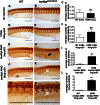
Motor axons are abnormal in Tardbpl-FL knocked down tardbpfh301/fh301embryos at 36 hpf. Lateral views of the whole-mounted tardbp+/+and tardbpfh301/fh301embryos stained with znp-1 to detect axons. (AC) Uninjected tardbp+/+(WT) and tardbpfh301/fh301 do not show any significant difference in axonal defects similar to control AMO (CoMo) injected groups (DF). (GI) Partial knockdown of Tardbpl-FL with tardbpl AMO +p53 results in a significant rise in the axonal defects in the tardbpfh301/fh301mutant zebrafish (P < 0.001). (J) Normal motor axons in tardbp+/+injected with tardbpl AMO 16ng. (K) Complete knockdown of tardbpl-FL in the tardbpfh301/fh301resulted in severe axonal outgrowth defects with complete arrest of axons at the horizontal myoseptum (P < 0.0001) (L). (M and N) Enlarged sections of (J and K) demonstrating severe axonal out growth defects in the double knockout (Tardbp and Tardbl-FL) embryos. Black arrow points out at an infrequent axonal truncation defect (low dose tardbpl AMO), while white arrows indicate numerous axonal out growth defects (high dose tardbpl AMO).
Figure 5.
Figure 5.
Alternative splicing of tardbpl gives rise to tardbpl-FL, which rescues the tardbpfh301/fh301 null phenotype. (A)Western blot of WT and tardbpfh301/fh301 embryos at 48 hpf, probed with h.TDP-43 Ab1, demonstrating relative over expression of Tardbpl-FL (solid black arrow) and the reduction in Tardbpl expression in the tardbpfh301/fh301 mutant zebrafish (asterisk). (B) Introns 5–6 of the tardbpl (ENSDART00000027255) on chromosome 23 contain a coding sequence, which potentially could give rise to a longer Tardbpl protein (Tardbpl-FL). (C) Predicted alternative splicing event, which results in inclusion of introns 5–6 creating tardbpl-FL. RT–PCR primers for common exon (exons 2 and 3), tardbpl-FL-specific primers (exons 4 and 5 but within intron 6 of tardbpl) and tardbpl-specific primer (exons 4 and 7) are also shown by arrows. (D) qRT–PCR estimation of expression of tardbpl and tardbpl-FL revealed a 2.42-fold increase (tardbpl-FL, ΔΔCT WT-tardbpfh301/fh301 of +1.27) in the tardbpl-FL expression in tardbpfh301/fh301mutant embryos compared with the WT embryos at 48 hpf and tardbpl expression was reduced in the tardbpfh301/fh301mutant embryos (tardbpl, ΔΔCT WT-tardbpfh301/fh301 of −0.213). (E) ClustalW2 alignment of the Tardbp and predicted Tardbpl-FL amino acid sequences. Both the N and the C termini are highly conserved. Tardbpl-FL is missing nine amino acids from the GRD, of which six are glycine residues (highlighted in grey).

References

    1. Ferraiuolo L., Kirby J., Grierson A.J., Sendtner M., Shaw P.J. Molecular pathways of motor neuron injury in amyotrophic lateral sclerosis. Nat. Rev. Neurol. 2011;7:616–630. - PubMed
    1. Lagier-Tourenne C., Polymenidou M., Cleveland D.W. TDP-43 and FUS/TLS, emerging roles in RNA processing and neurodegeneration. Hum. Mol. Genet. 2010;19:R46–R64. - PMC - PubMed
    1. Neumann M., Sampathu D.M., Kwong L.K., Truax A.C., Micsenyi M.C., Chou T.T., Bruce J., Schuck T., Grossman M., Clark C.M., et al. Ubiquitinated TDP-43 in frontotemporal lobar degeneration and amyotrophic lateral sclerosis. Science. 2006;314:130–133. - PubMed
    1. Geser F., Lee V.M., Trojanowski J.Q. Amyotrophic lateral sclerosis and frontotemporal lobar degeneration, a spectrum of TDP-43 proteinopathies. Neuropathology. 2010;30:103–112. - PMC - PubMed
    1. Wegorzewska I., Baloh R.H. TDP-43-based animal models of neurodegeneration, new insights into ALS pathology and pathophysiology. Neurodegener. Dis. 2011;8:262–274. - PMC - PubMed

Publication types

MeSH terms