. 2013 Feb 20;5(173):173ra24.
doi: 10.1126/scitranslmed.3004978.

Cross-hemispheric functional connectivity in the human fetal brain

Affiliations

Cross-hemispheric functional connectivity in the human fetal brain

Moriah E Thomason et al. Sci Transl Med. .

Abstract

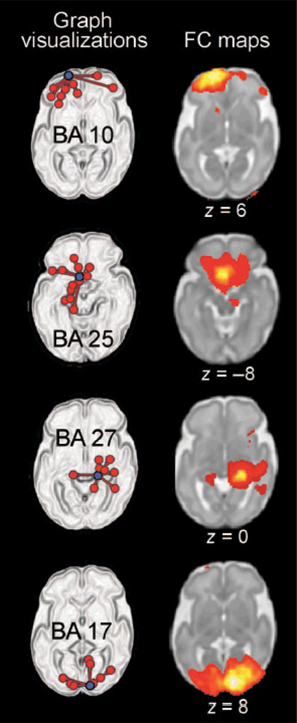
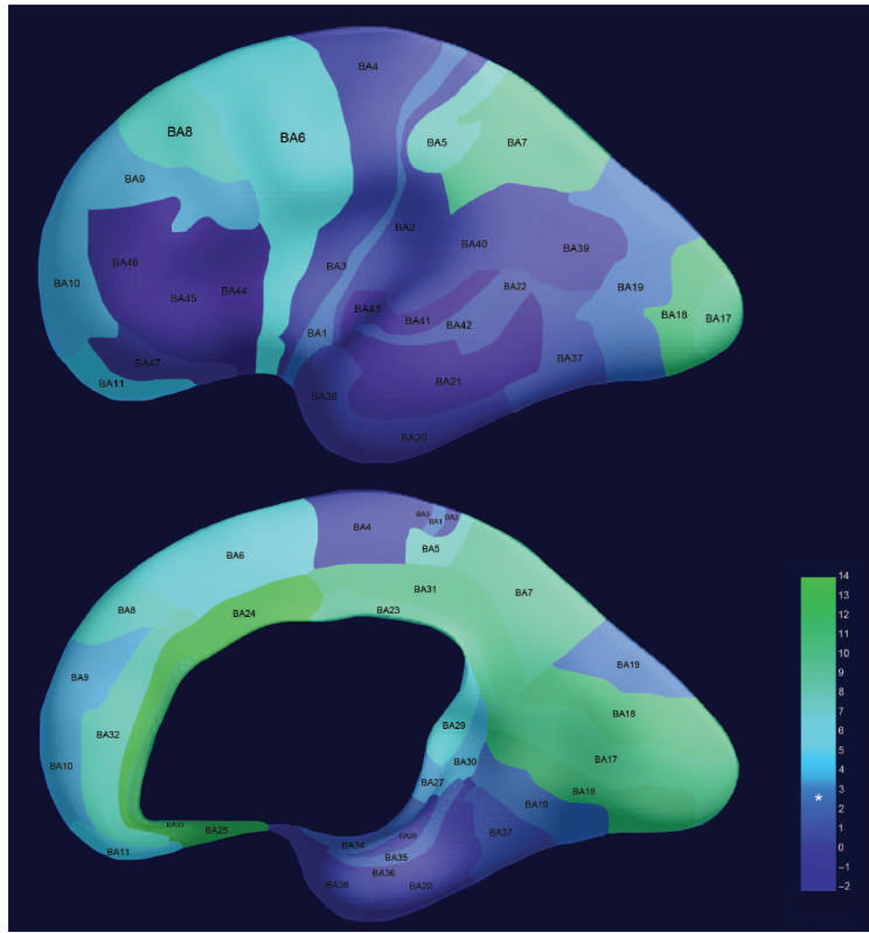
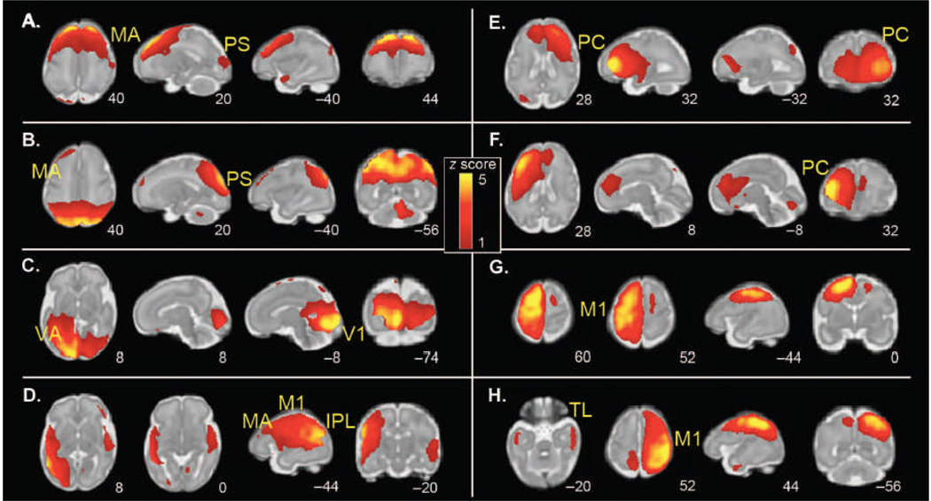
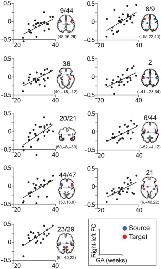
Compelling evidence indicates that psychiatric and developmental disorders are generally caused by disruptions in the functional connectivity (FC) of brain networks. Events occurring during development, and in particular during fetal life, have been implicated in the genesis of such disorders. However, the developmental timetable for the emergence of neural FC during human fetal life is unknown. We present the results of resting-state functional magnetic resonance imaging performed in 25 healthy human fetuses in the second and third trimesters of pregnancy (24 to 38 weeks of gestation). We report the presence of bilateral fetal brain FC and regional and age-related variation in FC. Significant bilateral connectivity was evident in half of the 42 areas tested, and the strength of FC between homologous cortical brain regions increased with advancing gestational age. We also observed medial to lateral gradients in fetal functional brain connectivity. These findings improve understanding of human fetal central nervous system development and provide a basis for examining the role of insults during fetal life in the subsequent development of disorders in neural FC.


Figures

Figure 1
Figure 1
Graph visualizations and FC maps of neural connectivity in a group of 25 fetuses. On the left, graph visualization depict a single “seed” region of interest (ROI) (in purple) along with all areas significantly functionally connected to that seed region (P < 0.05, corrected for multiple comparisons). The Brodmann’s area (BA) from which exemplar seeds are drawn is indicated on these graph depictions. FC maps on the right demonstrate the strength of connectivity (P < 0.001) from the same seed region that is depicted to the left but do so within a representative slice. Slice position is given below each FC map, where z = position in axial plane using Montreal Neurological Institute (MNI) coordinate space.
Figure 2
Figure 2
Regional differences in bilateral FC in 25 fetuses. Strength of right-left connectivity across the group is depicted as a color gradient with higher values corresponding to stronger FC. Asterisk is used to denote the statistical level (t = 2.85) at which measured FC exceeds a significance level of P (FDR) < 0.05. Colors are projected onto a three-dimensional (3D) rendering of the brain of a 30-week-old fetus. The lateral surface of the brain is shown in the top image, whereas the medial surface is shown in the bottom image.
Figure 3
Figure 3
Group ICA of spontaneous fMRI activity patterns in 25 fetuses (24 to 28 weeks of gestation). Sample axial, sagittal, and coronal slices corresponding to bilaterally represented independent components from ICA are overlaid onto the template of a 32-week fetal brain. The left side of the figure corresponds to the left side of the brain. Coordinates identifying slice locations are provided below each slice using the MNI coordinate space. ICA was used to derive a total of 14 maximally statistically independent brain networks, 8 of which were bilaterally distributed. (A to H) The following networks were detected: (A) motor association (MA) cortex; (B) peristriate (PS) cortex; (C) primary visual (V1) and visual association (VA) cortex; (D) inferior parietal lobule (IPL), primary motor (M1), and motor association cortex; (E) right frontal cortex; (F) left frontal cortex; (G) left primary motor cortex; and (H) right primary motor cortex and bilateral temporal lobe (TL).
Figure 4
Figure 4
Association between strength of bilateral FC and GA of fetuses. Scatter plots depict significant age effects across representative cortical areas. All correlation coefficients r are > 0.6 (Pearson; two-tailed) and P values are ≤ 0.001. Blue dots indicate sources used in SCA (seed) analyses. Red dots indicate approximate contralateral target areas where significant age-related increases in FC were observed. Target FC values and fetal age comprise the scatter plot data points. The precise FC coordinates of the target areas are included below cortical templates of a 32-week-old fetus, which are provided for visual reference. Bas are indicated above the reference images.
Figure 5
Figure 5
Fetal fMRI data preprocessing. Periods of time corresponding to low movement frames (shaded, gray background) were selected for each fetus and for each fun. Low movement frames were retained for further analysis. Masks were used to extract the fetal brain from surrounding tissue. Fetal fMRI brain data were then manually rotated (reoriented) into a standard position. Reoriented brain data were then processed using standard procedures for preparing functional MRI data for group analyses.

References

    1. Monk CS, Peltier SJ, Wiggins JL, Weng SJ, Carrasco M, Risi S, Lord C. Abnormalities of intrinsic functional connectivity in autism spectrum disorders. Neuroimage. 2009;47:764–772. - PMC - PubMed
    1. Weng SJ, Wiggins JL, Peltier SJ, Carrasco M, Risi S, Lord C, Monk CS. Alterations of resting state functional connectivity in the default network in adolescents with autism spectrum disorders. Brain Res. 2010;1313:202–214. - PMC - PubMed
    1. Cao Q, Zang Y, Sun L, Sui M, Long X, Zou Q, Wang Y. Abnormal neural activity in children with attention deficit hyperactivity disorder: A resting-state functional magnetic resonance imaging study. Neuroreport. 2006;17:1033–1036. - PubMed
    1. Tian L, Jiang T, Wang Y, Zang Y, He Y, Liang M, Sui M, Cao Q, Hu S, Peng M, Zhuo Y. Altered resting-state functional connectivity patterns of anterior cingulate cortex in adolescents with attention deficit hyperactivity disorder. Neurosci. Lett. 2006;400:39–43. - PubMed
    1. Bluhm RL, Miller J, Lanius RA, Osuch EA, Boksman K, Neufeld RW, Théberge J, Schaefer B, Williamson P. Spontaneous low-frequency fluctuations in the BOLD signal in schizophrenic patients: Anomalies in the default network. Schizophr. Bull. 2007;33:1004–1012. - PMC - PubMed

Publication types

LinkOut - more resources