. 2013 Feb 20;5(173):173ra25.
doi: 10.1126/scitranslmed.3005503.

Safety and efficacy of an injectable extracellular matrix hydrogel for treating myocardial infarction

Affiliations

Safety and efficacy of an injectable extracellular matrix hydrogel for treating myocardial infarction

Sonya B Seif-Naraghi et al. Sci Transl Med. .

Erratum in

  • Sci Transl Med. 2014 Apr 23;6(233):233er2

Abstract

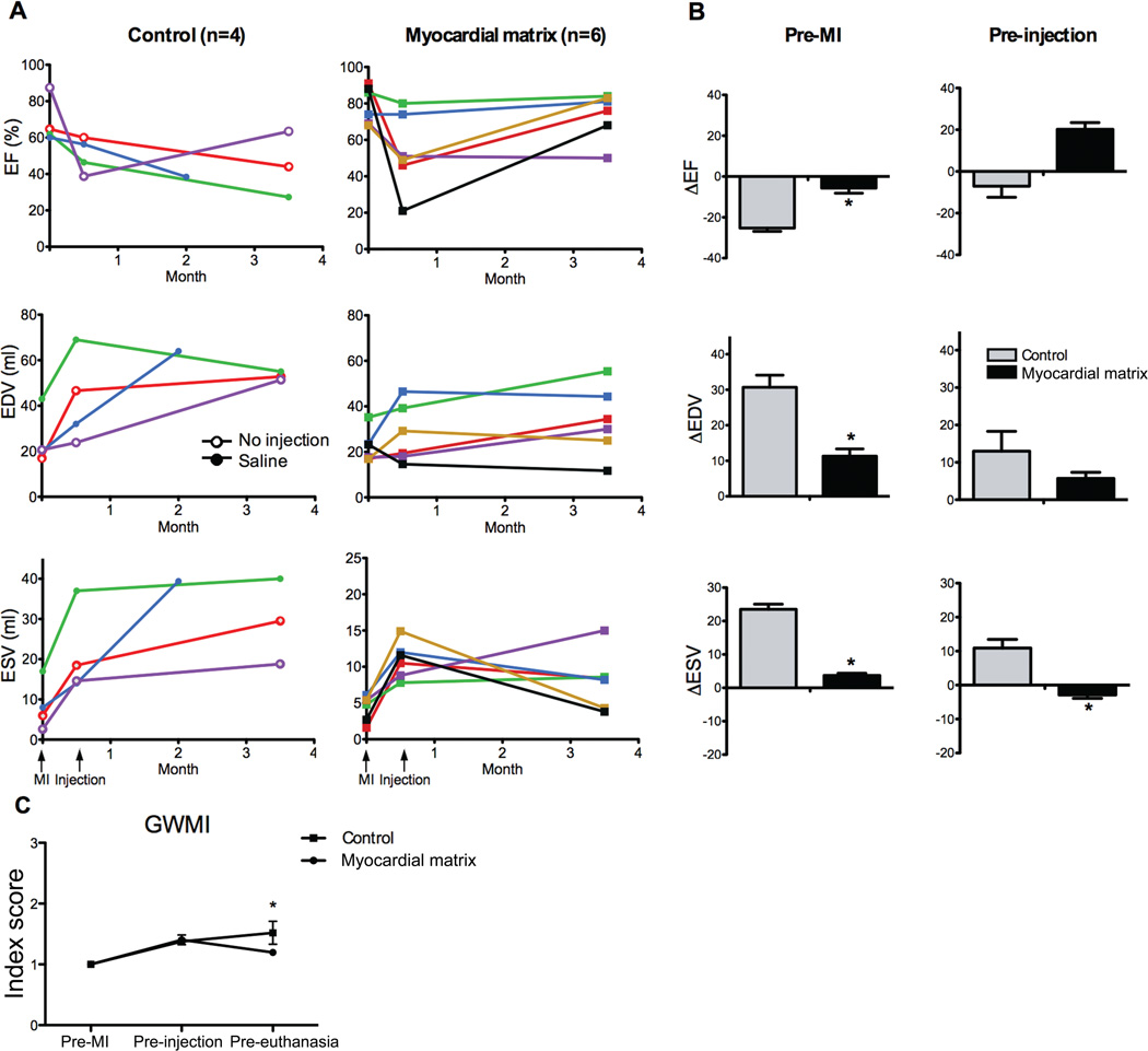
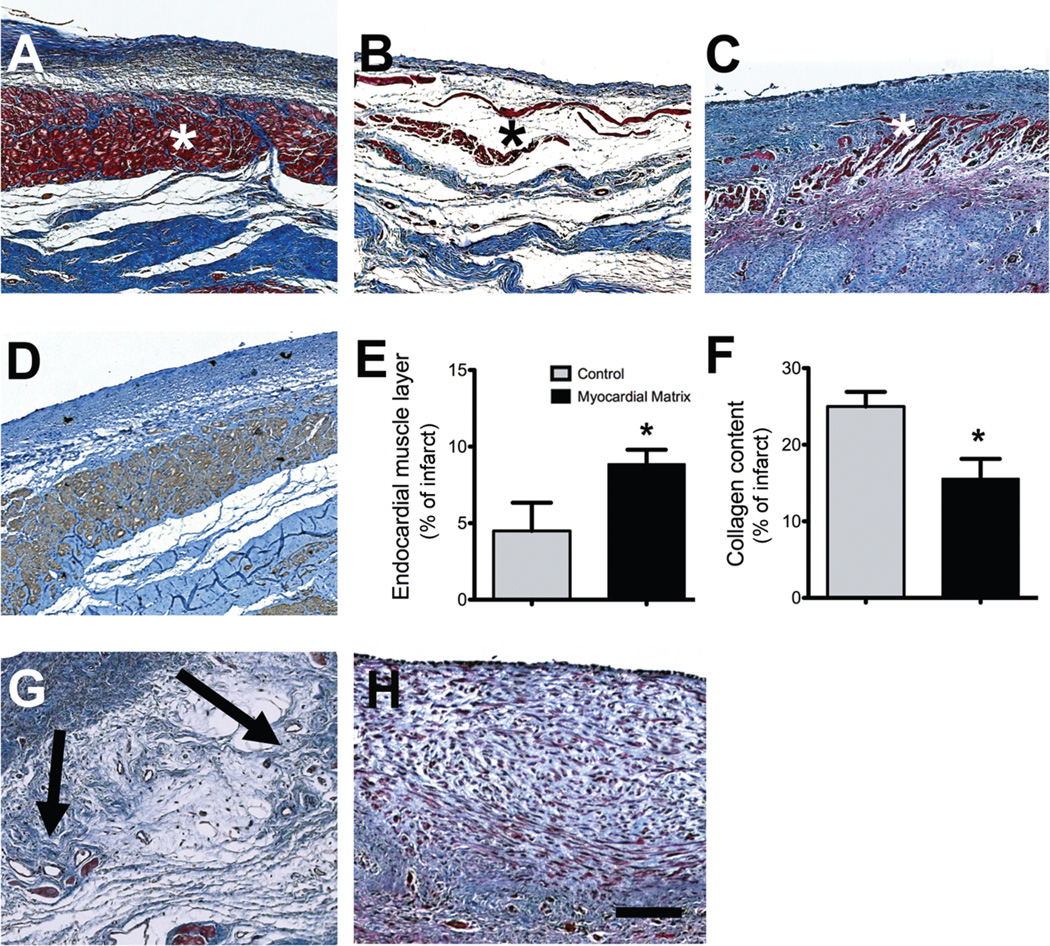
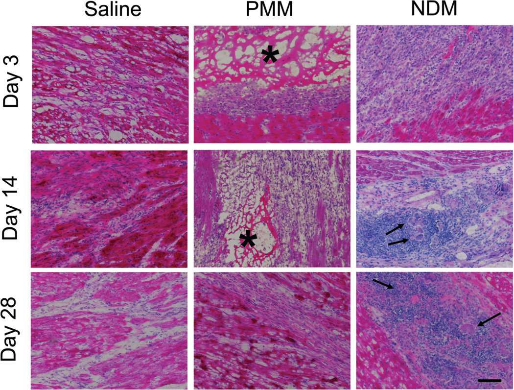
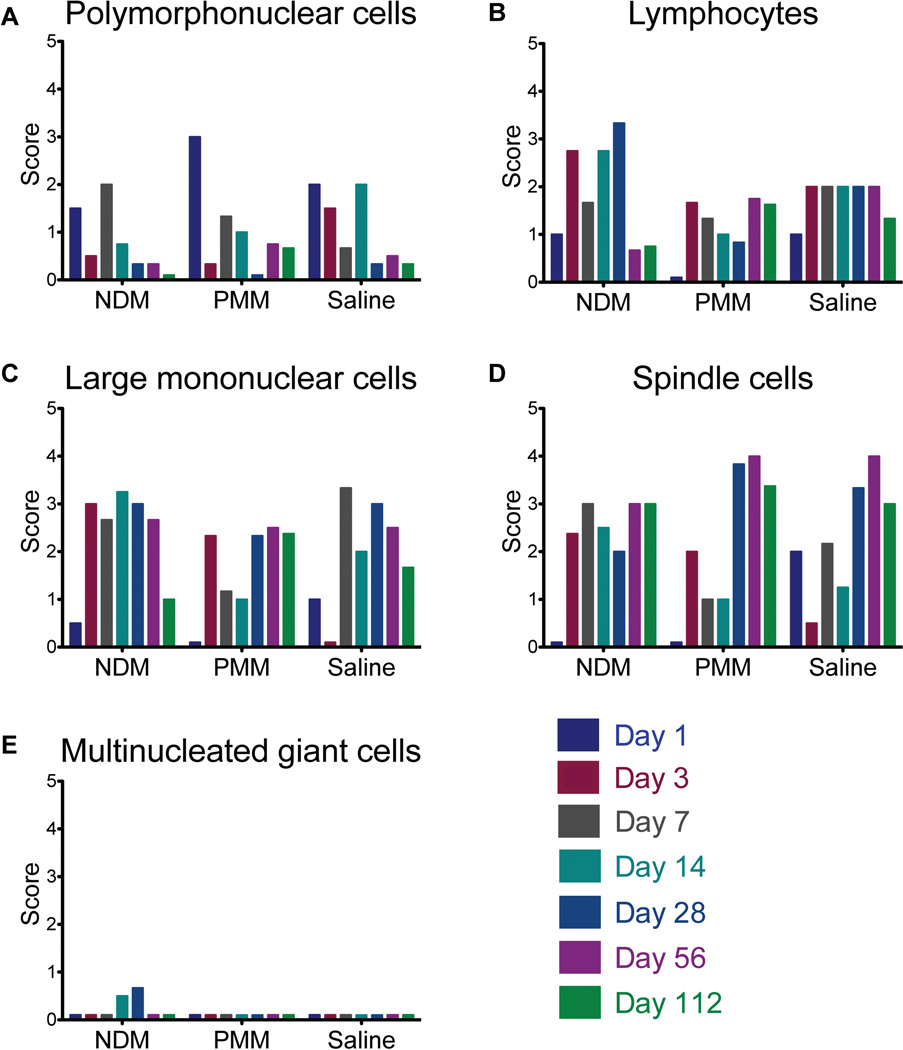
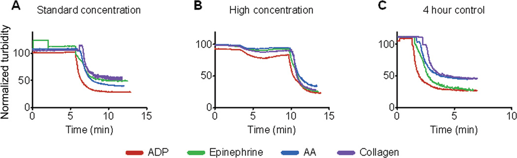
New therapies are needed to prevent heart failure after myocardial infarction (MI). As experimental treatment strategies for MI approach translation, safety and efficacy must be established in relevant animal models that mimic the clinical situation. We have developed an injectable hydrogel derived from porcine myocardial extracellular matrix as a scaffold for cardiac repair after MI. We establish the safety and efficacy of this injectable biomaterial in large- and small-animal studies that simulate the clinical setting. Infarcted pigs were treated with percutaneous transendocardial injections of the myocardial matrix hydrogel 2 weeks after MI and evaluated after 3 months. Echocardiography indicated improvement in cardiac function, ventricular volumes, and global wall motion scores. Furthermore, a significantly larger zone of cardiac muscle was found at the endocardium in matrix-injected pigs compared to controls. In rats, we establish the safety of this biomaterial and explore the host response via direct injection into the left ventricular lumen and in an inflammation study, both of which support the biocompatibility of this material. Hemocompatibility studies with human blood indicate that exposure to the material at relevant concentrations does not affect clotting times or platelet activation. This work therefore provides a strong platform to move forward in clinical studies with this cardiac-specific biomaterial that can be delivered by catheter.


Figures

Fig. 1
Fig. 1
Echocardiographic data shows improvement after injection of myocardial matrix. (A) Ejection fraction (EF), end-diastolic volume (EDV), and end-systolic volume (ESV) are shown for each animal pre-MI, pre-injection (2 weeks post-MI), and pre-euthanasia for both the control (n = 4) and matrix-injected (n = 6) groups. Control animals that received saline injections are indicated with open circles (green and blue lines); animals that received no injection are represented with open triangles (red and purple lines). (B) Comparison of the absolute changes in EF, EDV, and ESV from either pre-MI baseline or pre-injection to the time of euthanasia. (C) Global wall motion index improves in matrix-treated animals. Note that error bars are present but not visible for the matrix-injected group at pre-euthanasia. All data are means +/− SEM. *p < 0.05 versus control (Student’s t-test).
Fig. 2
Fig. 2
NOGA mapping and infarct expansion. (A) Representative unipolar area NOGA maps taken at time of injection and euthanasia. Images are thresholded to show the cutoff (6.9 mV) for the infarct area. (B) Infarct expansion calculated by unipolar area in myocardial matrix treated animals (n = 6) compared to the control group (n = 2). (C) Infarct expansion calculated via linear local shortening. Data are means +/− SEM. *p < 0.05 (Student’s t-test).
Fig. 3
Fig. 3
Histological characterization of infarcted pig hearts. (A to C) Masson’s trichrome staining images are representative of 6 matrix-injected pigs and 4 control animals. (A) Matrix-injected hearts had a distinct, thick endocardium (red, indicated with an asterisk). (B) Non-injected control animals had a loose fibrillar layer (blue) beneath the endothelium. (C) In saline-injected control animals, the endocardium was moderately thickened (red) (D) An adjacent tissue section for the matrix-injected animal in (A) stained for cardiac troponin-T, indicating the presence of cardiomyocytes. (E) Area of endocardial layer of muscle as a proportion of the infarct. (F) Percentage of collagen content in the infarcts. Data are means +/− SEM and were obtained from Masson’s trichrome slides (A to C). *p < 0.05, (Student’s t-test). (G and H) Matrix-injected hearts contained foci of neovascularization in the area below the endocardium (G, arrows) but none of the saline or non-injected control hearts showed these areas of neovascularization (H). Scale bar is 200 µm.
Fig. 4
Fig. 4
Cell infiltration in matrix-injected rat hearts. Representative histological sections from rat hearts injected with saline, porcine myocardial matrix (PMM), or non-decellularized matrix (NDM) at days 3, 14, and 28. Inflammation and multinucleated giant cells are present in the NDM groups at days 14 and 28 (arrows). The PMM (asterisk-marked porous network) was completely degraded by 28 days. Scale bar is 200 µm.
Fig. 5
Fig. 5
Matrix biocompatibility assessment in rats. (A to E) Average score for infiltration and degree of inflammation with respect to polymorphonuclear cells (A), mononuclear cells (B), lymphocytes (C), spindle cells (fibroblasts, myofibroblasts, and cardiomyocytes) (D), and multinucleated giant cells (E). On this scale, 0 = absent, 1 = minimal, 2 = mild, 3 = moderate, and 4 = marked. Due to freezing artifact, some tissue could not be analyzed and the data shown here is a composite score from 2 or 3 hearts per time point per group. .
Fig. 6
Fig. 6
Platelet activation. (A and B) Addition of myocardial matrix to human platelet-richplasma was done at a standard concentration (1:10,000) (A) and a high concentration (1:2500) (B) and the samples were evaluated for at least five minutes. After the samples plateaued, agonists were added (at 10 minutes). (C) A control was performed after four hours to ensure normal activity of the samples and validity of the data in response to agonists. Data shown are representative traces from 4 repeated experiments.

References

    1. Roger VL, et al. Heart disease and stroke statistics--2012 update: a report from the American Heart Association. Circulation. 2012 Jan 3;125:e2. - PMC - PubMed
    1. Christman KL, Lee RJ. Biomaterials for the treatment of myocardial infarction. J Am Coll Cardiol. 2006 Sep 5;48:907. - PubMed
    1. Rane AA, Christman KL. Biomaterials for the treatment of myocardial infarction a 5-year update. Journal of the American College of Cardiology. 2011 Dec 13;58:2615. - PubMed
    1. Nelson DM, Ma Z, Fujimoto KL, Hashizume R, Wagner WR. Intra-myocardial biomaterial injection therapy in the treatment of heart failure: Materials, outcomes and challenges. Acta Biomater. 2011 Jan;7:1. - PMC - PubMed
    1. Johnson TD, Christman KL. Injectable hydrogel therapies and their delivery strategies for treating myocardial infarction. Expert opinion on drug delivery. 2012 Nov 9; - PubMed

Publication types