. 2013 Apr;24(8):1134-52.
doi: 10.1091/mbc.E12-11-0801. Epub 2013 Feb 20.

The N-DRC forms a conserved biochemical complex that maintains outer doublet alignment and limits microtubule sliding in motile axonemes

Affiliations

The N-DRC forms a conserved biochemical complex that maintains outer doublet alignment and limits microtubule sliding in motile axonemes

Raqual Bower et al. Mol Biol Cell. 2013 Apr.

Abstract

The nexin-dynein regulatory complex (N-DRC) is proposed to coordinate dynein arm activity and interconnect doublet microtubules. Here we identify a conserved region in DRC4 critical for assembly of the N-DRC into the axoneme. At least 10 subunits associate with DRC4 to form a discrete complex distinct from other axonemal substructures. Transformation of drc4 mutants with epitope-tagged DRC4 rescues the motility defects and restores assembly of missing DRC subunits and associated inner-arm dyneins. Four new DRC subunits contain calcium-signaling motifs and/or AAA domains and are nearly ubiquitous in species with motile cilia. However, drc mutants are motile and maintain the 9 + 2 organization of the axoneme. To evaluate the function of the N-DRC, we analyzed ATP-induced reactivation of isolated axonemes. Rather than the reactivated bending observed with wild-type axonemes, ATP addition to drc-mutant axonemes resulted in splaying of doublets in the distal region, followed by oscillatory bending between pairs of doublets. Thus the N-DRC provides some but not all of the resistance to microtubule sliding and helps to maintain optimal alignment of doublets for productive flagellar motility. These findings provide new insights into the mechanisms that regulate motility and further highlight the importance of the proximal region of the axoneme in generating flagellar bending.


Figures

FIGURE 1:
FIGURE 1:
DRC4 is truncated by alternative splicing in sup-pf3. (A) Western blot of WT and mutant axonemes probed with antibodies to DRC4 reveals the presence of truncated DRC4 subunits in sup-pf3. Anti-Rib43 serves as a loading control. The outer-arm subunit IC2 is reduced in the outer-arm mutants pf28 and sup-pf2. DRC2 and tektin are reduced only in pf3. (B) Clustal W alignment of the human GAS11 and Chlamydomonas DRC4 polypeptide sequences. The deleted amino acids (outlined in the black box) correspond to a region that is highly conserved between DRC4 orthologues. (C) Predicted structural domains in WT and sup-pf3 DRC4 subunits. The deleted regions are predicted to alter the arrangement of coiled-coil domains 1 and 2 in DRC4.
FIGURE 2:
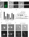
FIGURE 2:
PF2-GFP rescues the mutant phenotypes associated with pf2 and sup-pf3. (A) Live imaging of an immobilized PF2-GFP rescued cell by DIC and fluorescence microscopy reveals that DRC4-GFP is located near the basal body region and along the length of the flagella. (B) DIC and fluorescence imaging of fixed PF2-GFP rescued cells (left) and mutant cells (right, pf2-4 and sup-pf3) shows that the GFP signal is observed exclusively in the rescued strains. (C) The forward swimming velocities of WT, mutant, and rescued strains were measured by phase contrast microscopy. The total number of cells measured for each strain (n) is indicated. Both mutants were significantly slower than WT (p < 0.005). Rescued strains were faster than the mutant strains but not completely WT (p < 0.005). The sup-pf3::PF2-GFP rescued strains were also slightly but significantly slower than pf2::PF2-GFP (p < 0.05). (D) A Western blot of isolated axonemes was probed with antibodies against the DRC4 fusion protein antibody (top) and the RSP16 subunit (bottom). Truncated DRC4 subunits are found in sup-pf3 and two rescued strains (left), whereas a larger, more abundant band at ∼75 kDa is observed in the PF2-GFP rescued strains. (E) DIC and fluorescence images of dikaryons fixed at 10 and 30 min after mixing WT and pf2-4::PF2-GFP cells and stained with an antibody against GFP. No significant accumulation of the GFP signal was observed in the WT flagella. (F) Images of dikaryons fixed at 10 and 60 min after mixing pf2-1 and pf2-4::PF2-4-GFP cells and stained with the GFP antibody. Accumulation of GFP signal was initially observed at the flagellar tips and gradually spread to the proximal region. Scale bars, 5 μm.
FIGURE 3:
FIGURE 3:
Inner-arm dynein subspecies in pf2. (A) Isolated axonemes from WT, pf2-4, and pf2-4::PF2-HA were separated on a 5% SDS–PAGE gel, the DHC regions were excised, and three to five replicates were analyzed by MS/MS and spectral counting. The total number of spectra for each DHC subspecies was expressed as a percentage of the total number of spectra for the two DHCs of the I1 dynein. DHC8 was substantially reduced in pf2, and DHC2 was slightly reduced (p <0.005). (B) Western blot of WT and pf2 axonemes probed with antibodies against various dynein subunits. No obvious defects were observed with antibodies against the outer-arm (OA) subunit IC69 or the inner-arm subunits shown here. No antibodies were available for DHC2 or DHC8.
FIGURE 4:
FIGURE 4:
Identification of polypeptides that interact with DRC4. (A) Silver-stained gel of immunoprecipitates obtained with an HA antibody and extracts from pf2 and PF2-HA axonemes. Although several polypeptides were nonspecifically bound to HA beads in both samples, several unique bands were observed in the PF2-HA sample. See Table 1 for polypeptides identified by MS/MS. (B) Diagrams showing predicted polypeptide domains in the candidate DRC subunits identified by coimmunoprecipitation and/or iTRAQ analyses. (C) Western blot of WT, pf2, sup-pf3, and sup-pf4 axonemes probed with antibodies against candidate DRC subunits and other axonemal polypeptides. Several subunits identified as missing in pf2 were also missing or reduced in sup-pf3. Only FAP155/DRC5 was missing in sup-pf4. Antibodies against subunits of the I1 dynein (IC140), CSC (FAP61/CaM-IP3), RS (RSP16), and FAP59/CCDC39 serve as loading controls. (D) Western blots of isolated axonemes (WA) and extracts obtained by sequential treatment with 0.6 M NaCl (HS), 0.2, 0.4, and 0.6 M NaI, and the final extracted outer doublets (OD). DRC subunits were coextracted from WT and PF2-HA with 0.4–0.6 M NaI, but some subunits were more readily extracted from sup-pf3 at lower ionic strengths.
FIGURE 5:
FIGURE 5:
Sucrose density gradient centrifugation identifies subcomplexes within the DRC. Western blots of fractions obtained by analyzing DRC-containing extracts on 5–20% sucrose density gradients. The bottoms (20% sucrose) of the gradients are on the left. (A) PF2-HA, all DRC subunits cosediment near the bottom of the gradient; (B) sup-pf4, DRC subunits dissociate into multiple peaks; (C) sup-pf3, DRC subunits form three distinct subcomplexes; (D) SUP-PF4-HA, most DRC subunits reassociate to form a large complex that sediments near the bottom of the gradient.
FIGURE 6:
FIGURE 6:
The N-DRC maintains alignment between outer doublet microtubules in the presence of ATP. (A) Responses of full-length WT or mutant axonemes upon addition of 0.1 mM ATP or 0.1 mM ATP and protease. (B) Quantification of the numbers of axonemes that remained intact, splayed at their distal ends, or underwent sliding disintegration in the presence of 0.1 mM ATP alone. The majority of WT axonemes remained intact (and occasionally were observed to reactivate flagellar beating), whereas axonemes from drc-mutant strains splayed at their distal ends. Axonemes from rescued strains remained intact. Scale bar is 4 µm.

References

    1. Ahmed NT, Gao C, Lucker BF, Cole DG, Mitchell DR. ODA16 aids axonemal outer row dynein assembly through an interaction with the intraflagellar transport machinery. J Cell Biol. 2008;183:313–322. - PMC - PubMed
    1. Akkina SK, Zhang Y, Nelsestuen GL, Oetting WS, Ibrahlm HN. Temporal stability of the urinary proteome after kidney transplant: more sensitive than protein composition. J Proteome Res. 2009;8:94–103. - PMC - PubMed
    1. Andersen JD, Boylan KL, Xue FS, Anderson LB, Witthuhn BA, Markowski TW, Higgins L, Skubitz AP. Identification of candidate biomarkers in ovarian cancer serum by depletion of highly abundant proteins and differential in-gel electrophoresis. Electrophoresis. 2010;31:599–610. - PMC - PubMed
    1. Aoyama S, Kamiya R. Cyclical interactions between two outer doublet microtubules in split flagellar axonemes. Biophys J. 2005;89:3261–3268. - PMC - PubMed
    1. Armbrust EV, et al. The genome of the diatom Thalassiosira pseudonana: ecology, evolution, and metabolism. Science. 2004;306:79–86. - PubMed

Publication types

LinkOut - more resources