. 2013 May 17;8(5):958-66.
doi: 10.1021/cb3005726. Epub 2013 Mar 7.

Evidence for context-dependent complementarity of non-Shine-Dalgarno ribosome binding sites to Escherichia coli rRNA

Affiliations

Evidence for context-dependent complementarity of non-Shine-Dalgarno ribosome binding sites to Escherichia coli rRNA

Pamela A Barendt et al. ACS Chem Biol. .

Abstract

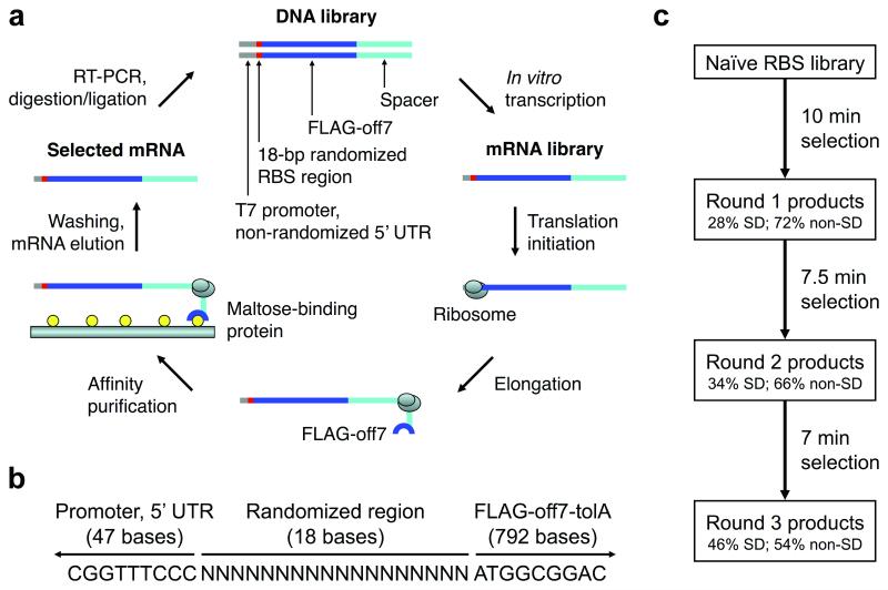
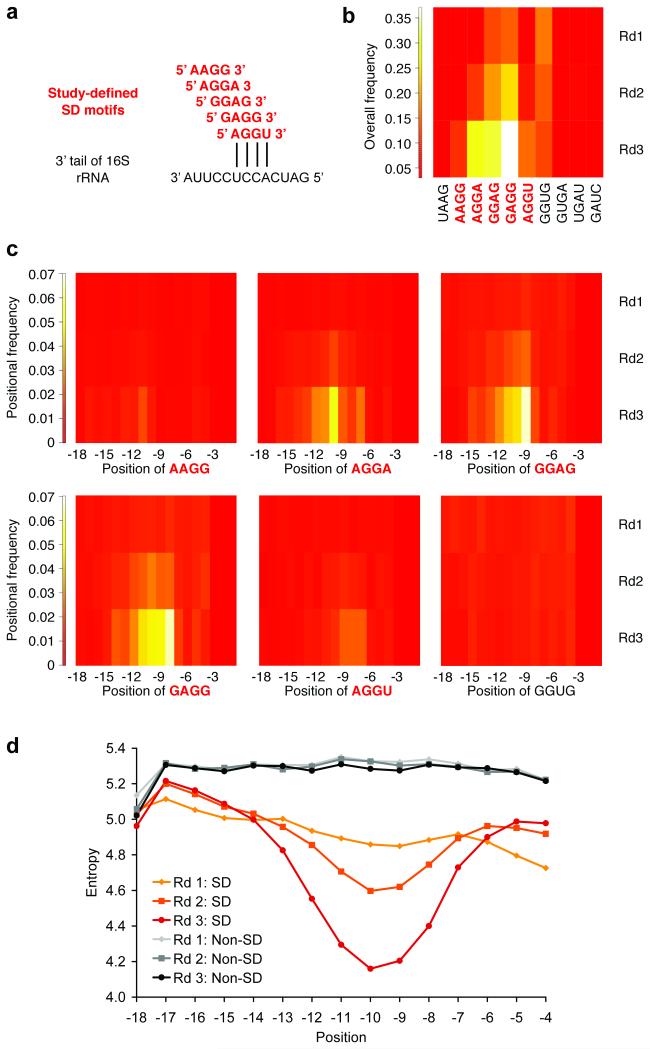
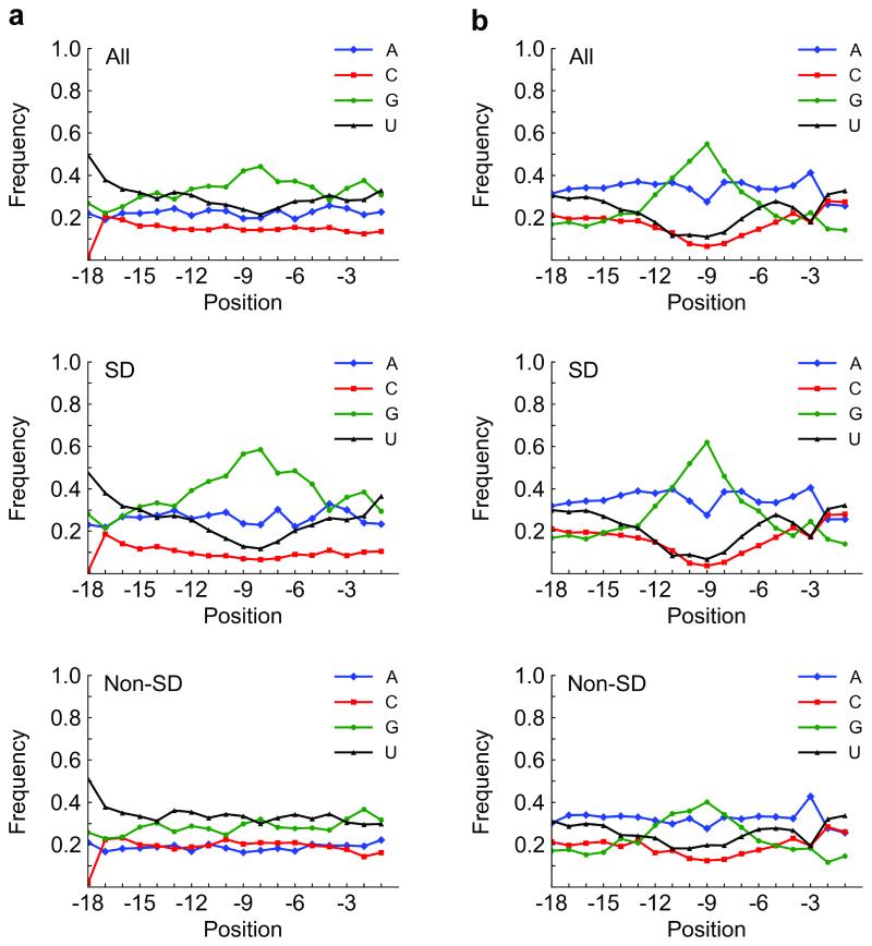
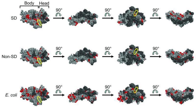
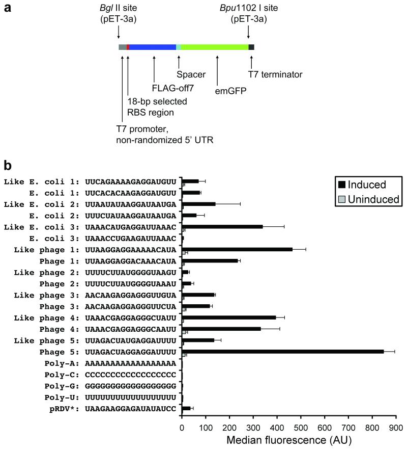
While the ribosome has evolved to function in complex intracellular environments, these contexts do not easily allow for the study of its inherent capabilities. We have used a synthetic, well-defined Escherichia coli (E. coli)-based translation system in conjunction with ribosome display, a powerful in vitro selection method, to identify ribosome binding sites (RBSs) that can promote the efficient translation of messenger RNAs (mRNAs) with a leader length representative of natural E. coli mRNAs. In previous work, we used a longer leader sequence and unexpectedly recovered highly efficient cytosine-rich sequences with complementarity to the 16S ribosomal RNA (rRNA) and similarity to eukaryotic RBSs. In the current study, Shine-Dalgarno (SD) sequences were prevalent, but non-SD sequences were also heavily enriched and were dominated by novel guanine- and uracil-rich motifs that showed statistically significant complementarity to the 16S rRNA. Additionally, only SD motifs exhibited position-dependent decreases in sequence entropy, indicating that non-SD motifs likely operate by increasing the local concentration of ribosomes in the vicinity of the start codon, rather than by a position-dependent mechanism. These results further support the putative generality of mRNA-rRNA complementarity in facilitating mRNA translation but also suggest that context (e.g., leader length and composition) dictates the specific subset of possible RBSs that are used for efficient translation of a given transcript.


Figures

Figure 1
Figure 1
Ribosome display method, library context, and selection scheme. a) In our adaptation of ribosome display for the selection of efficiently translated sequences, the naïve DNA library contained an 18-bp randomized RBS region before the start codon. Selection pressure was increased over multiple rounds by progressively limiting the time of in vitro translation. b) The DNA context of the randomized RBS region is shown. The T7 promoter and 5′ UTR stem-loop derived from the ribosome display vector, pRDV, are upstream (47 bases in the DNA construct, 21 bases in the mRNA transcript). The coding region (downstream) contains a fusion protein with a FLAG tag, Off7 (a designed ankyrin repeat protein which binds maltose-binding protein), TolA (an unstructured spacer derived from E. coli tolA which allows Off7 to exit the ribosomal tunnel and fold properly), and no stop codon. c) The selection scheme is shown. The naïve RBS library was subjected to three selection rounds of increasing stringency: 10 min, 7.5 min, and 7 min translation. SD sequences were enriched between rounds, but many non-SD sequences remained in the pool after three rounds. Adapted from (8). 5′ UTR, 5′ untranslated region; RBS, ribosome binding site; SD, Shine-Dalgarno.
Figure 2
Figure 2
Enrichment of SD sequences. a) The alignment of study-defined SD motifs (red) with the 3′ tail of the 16S rRNA (black) is shown. b) Overall enrichment of SD sequences over three rounds (Rd1, Rd2, and Rd3) is shown. For comparison, we present all 10 four-base subsets of the reverse complement (5′-UAAGGAGGUGAUC-3′) to the 13 unpaired bases at the 3′ end of the 16S rRNA (5′-GAUCACCUCCUUA-3′) in our selected sequences: UAAG, AAGG, AGGA, GGAG, GAGG, AGGU, GGUG, GUGA, UGAU, and GAUC. c) All study-defined SD motifs (AAGG, AGGA, GGAG, GAGG, and AGGU) exhibited position-dependent enrichment according to their alignment with the 16S rRNA. GGUG (also shown) was enriched overall, but not in a position-dependent manner. d) The position-dependent entropy of four-base windows of 18-base sequences with or without an SD motif over three rounds is shown. Similar plots were observed for five-, six-, seven-, and eight-base windows. Position is specified by the first base of the motif relative to the start codon. SD, Shine-Dalgarno.
Figure 3
Figure 3
Base content vs. position in our selection and in E. coli. a) Base content vs. position in all, SD (46%), and non-SD (54%) sequences, respectively, in our selection (after third round). Overall (top panel), the nucleotide G is abundant around position −9 or −8, which reflects SD content (middle panel). Interestingly, the non-SD sequences (bottom panel) are mostly G/U-rich. b) Base content vs. position in all, SD (67%), and non-SD (33%) sequences, respectively, from the 18 bases before the start codon in E. coli K12 W3110 (NCBI TaxID 316407; Transterm database (22)). Generally, the base distribution resulting from our selections mimicked that in E. coli. The G peak that appears in our library appears at approximately the same position in E. coli, primarily from the SD sequence. Position is specified by the first base of the motif relative to the start codon. SD, Shine-Dalgarno.
Figure 4
Figure 4
Distribution of potential sites for base-pairing of RBSs (selected library [SD only], top row; selected library [non-SD only], middle row; and E. coli, bottom row) to 16S rRNA. Regions on the E. coli 30S ribosomal subunit with significant complementarity to the RBS population of interest (p-value < 0.01; Bonferroni-corrected) were determined. Significant six-base windows that shared five bases with at least one neighboring significant window are highlighted in red (PyMOL rendering of PDB 3DF1). Four different views convey the general distribution of these potential base-pairing sites over the small ribosomal subunit. The first view in each row shows the face that becomes buried after assembly with the large ribosomal subunit. The approximate position of the anti-SD sequence is indicated by the yellow ellipse. 16S rRNA = light gray; ribosomal proteins = dark gray. RBS, ribosome binding site; SD, Shine-Dalgarno.
Figure 5
Figure 5
In vivo expression. a) Expression cassettes containing an RBS followed by FLAG-off7-emGFP were cloned into pET-3a and expressed in BL21(DE3)pLysS. b) Average median fluorescence of each clone is shown. Error bars indicate standard deviation of at least three experiments. Library members having maximal similarity to E. coli or phage 5′ UTRs and the corresponding natural sequences are shown, along with the four homopolymers (negative controls) and the 18 bases before the start codon in the ribosome display vector, pRDV (* indicates that these 18 bases are situated in the short leader context). 5′ UTR, 5′ untranslated region; emGFP, Emerald GFP; RBS, ribosome binding site.

References

    1. Shine J, Dalgarno L. The 3′-terminal sequence of Escherichia coli 16S ribosomal RNA: complementarity to nonsense triplets and ribosome binding sites. Proc. Natl. Acad. Sci. U. S. A. 1974;71:1342–1346. - PMC - PubMed
    1. Chang B, Halgamuge S, Tang S-L. Analysis of SD sequences in completed microbial genomes: non-SD-led genes are as common as SD-led genes. Gene. 2006;373:90–99. - PubMed
    1. Kozak M. An analysis of 5′-noncoding sequences from 699 vertebrate messenger RNAs. Nucleic Acids Res. 1987;15:8125–8148. - PMC - PubMed
    1. Cavener DR, Ray SC. Eukaryotic start and stop translation sites. Nucleic Acids Res. 1991;19:3185–3192. - PMC - PubMed
    1. Gingold H, Pilpel Y. Determinants of translation efficiency and accuracy. Mol. Syst. Biol. 2011;7:481. - PMC - PubMed

Publication types

LinkOut - more resources