Review
. 2013 Mar;13(3):260-8.
doi: 10.1016/S1473-3099(13)70001-X.

Immunosuppression in sepsis: a novel understanding of the disorder and a new therapeutic approach

Affiliations
Review

Immunosuppression in sepsis: a novel understanding of the disorder and a new therapeutic approach

Richard S Hotchkiss et al. Lancet Infect Dis. 2013 Mar.

Abstract

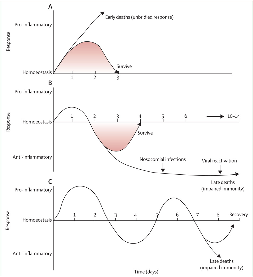
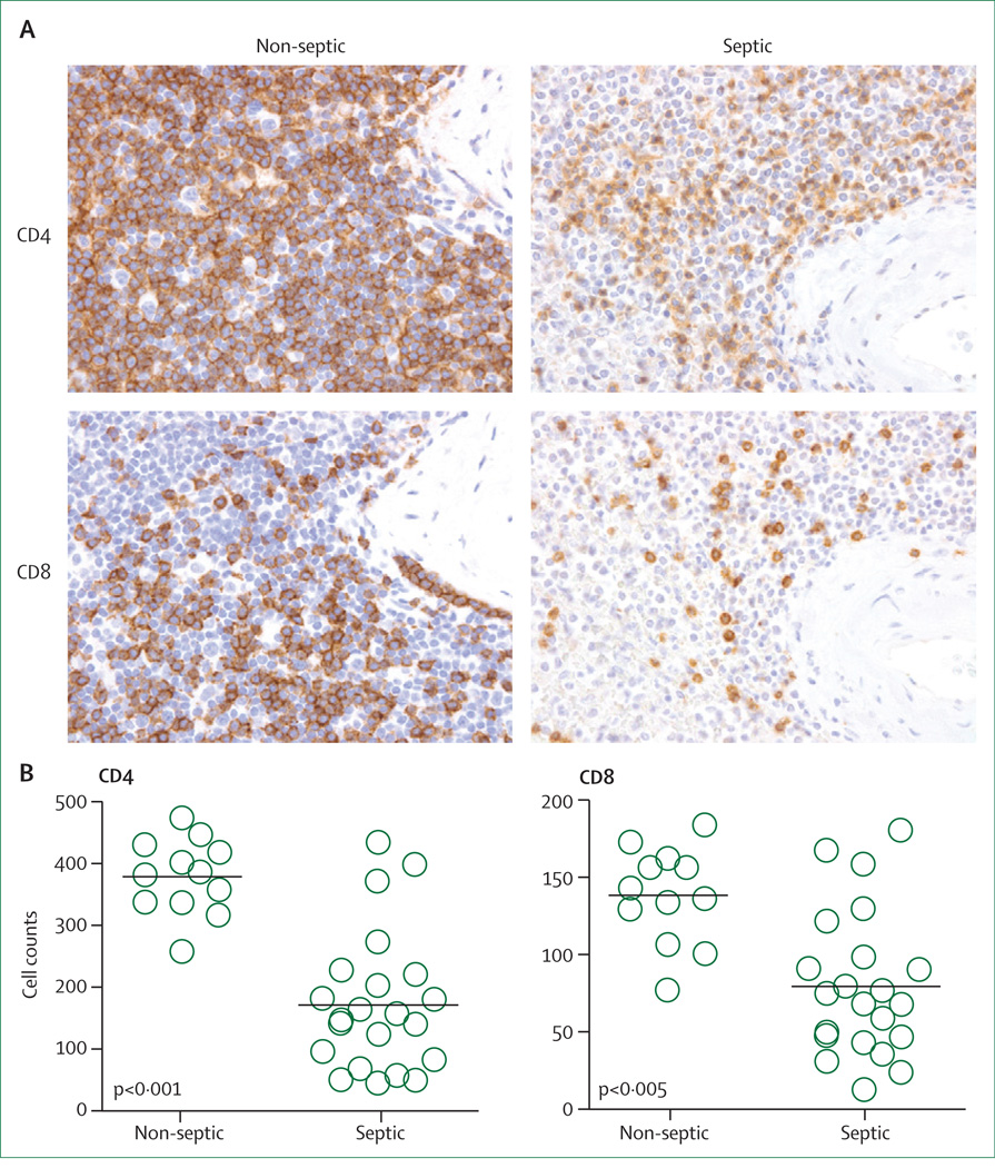
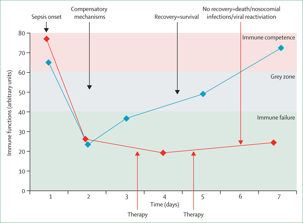
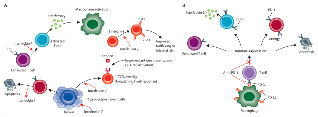
Failures of highly touted trials have caused experts to call for re-evaluation of the current approach toward sepsis. New research has revealed key pathogenic mechanisms; autopsy results have shown that most patients admitted to intensive care units for treatment of sepsis had unresolved septic foci at post mortem, suggesting that patients were unable to eradicate invading pathogens and were more susceptible to nosocomial organisms, or both. These results suggest that therapies that improve host immunity might increase survival. Additional work showed that cytokine production by splenocytes taken post mortem from patients who died of sepsis is profoundly suppressed, possibly because of so-called T-cell exhaustion-a newly recognised immunosuppressive mechanism that occurs with chronic antigenic stimulation. Results from two clinical trials of biomarker-guided therapeutic drugs that boosted immunity showed promising findings in sepsis. Collectively, these studies emphasise the degree of immunosuppression that occurs in sepsis, and explain why many previous sepsis trials which were directed at blocking inflammatory mediators or pathogen recognition signalling pathways failed. Finally, highly encouraging results from use of the new immunomodulatory molecules interleukin 7 and anti-programmed cell death 1 in infectious disease point the way for possible use in sepsis. We hypothesise that immunoadjuvant therapy represents the next major advance in sepsis.


Figures

Figure 1
Figure 1. Potential inflammatory responses in sepsis
Immune responses in sepsis are determined by many factors including pathogen virulence, size of bacterial inoculum, comorbidities, etc. (A) Although both proinflammatory and anti-inflammatory responses begin rapidly after sepsis, the initial response in previously healthy patients with severe sepsis is typified by an overwhelming hyperinflammatory phase with fever, hyperdynamic circulation, and shock. Deaths in this early phase of sepsis are generally due to cardiovascular collapse, metabolic derangements, and multiple organ dysfunction. Although no particular anti-inflammatory therapies have improved survival in large phase 3 trials, short acting anti-inflammatory or anticytokine therapies offer a theoretical benefit. (B) Many patients who develop sepsis are elderly with numerous comorbidities that impair immune response. When these individuals develop sepsis, a blunted or absent hyperinflammatory phase is common, and patients rapidly develop impaired immunity and an anti-inflammatory state. Immunoadjuvant therapy that boosts immunity offers promise in this setting. (C) A third theoretical immunological response to sepsis is characterised by cycling between hyperinflammatory and hypoinflammatory states. According to this theory, patients who develop sepsis have an initial hyperinflammatory response followed by a hypoinflammatory state. With the development of a new secondary infection, patients have a repeat hyperinflammatory response and may either recover or re-enter the hypoinflammatory phase. Patients can die in either state. There is less evidence for this theory, and the longer the sepsis continues the more likely a patient is to develop profound immunosuppression.
Figure 2
Figure 2. Depletion of splenic lymphocytes in septic patients
(A) Spleens from patients with or without sepsis were obtained by rapid post-mortem sampling and immunostained for CD4, or CD8 T cells. An investigator blinded to sample identity examined the slides. CD4 and CD8 T cells are brown in colour (400× magnification). (B) CD4 and CD8 T cells are decreased in patients with sepsis relative to control patients without sepsis. Cell counts for CD4 and CD8 T cells obtained by counting the number of cells or field in periarteriolar lymphoid sheaths. N=12 non-septic and N=22 septic. Figure modified with permission from the American Medical Association.
Figure 3
Figure 3. Immunostimulation therapy in sepsis: a new approach
New biomarker-based methods to semi-quantitate the degree of immunosuppression in septic patients are now being used. For example, flow cytometric quantitation of circulating blood monocyte expression of HLA-DR has been used to identify patients who would respond to granulocyte macrophage colony stimulating factor (GM-CSF). In the future, other biomarkers that are currently used in cancer immunotherapy will probably be used. Monocyte expression of programmed cell-death ligand-1 (PD-L1) could be used to guide therapy with anti-PD-1 antibody. Patients who have persistently low absolute lymphocyte counts could be candidates for interleukin-7 therapy. Patients with infections caused by weakly virulent pathogens including Candida spp are also candidates for immunotherapy. Therapy refers to immunostimulation for most severely immunodepressed patients, identified via immunomonitoring.
Figure 4
Figure 4. Interleukin 7 and anti-PD-1 immunotherapy in sepsis
Interleukin 7 (A) acts to reverse immunosuppression by multiple mechanisms including increased production of CD4 and CD8 T cells, blockade of sepsis-induced apoptosis, reversal of T-cell exhaustion, increased interferon γ production leading to macrophage activation, increased integrin expression leading to improved T-cell recruitment to infected areas, and increased T-cell receptor (TCR) diversity. Anti-PD-1 antibody (B) will prevent interaction of programmed cell-death ligand-1 (PD-L1), which is expressed on macrophages with PD-1 receptor, which is expressed on T cells. Thus, anti-PD-1 antibody will prevent formation of exhausted T cells, decrease interleukin 10 production, prevent T-cell anergy, and decrease sepsis-induced apoptosis. LFA=leucocyte function-associated antigen. VLA=very late antigen. PD-1=programmed cell death 1.

Comment in

  • Adjunctive treatment of severe sepsis.
    Dinubile M. Dinubile M. Lancet Infect Dis. 2013 Nov;13(11):917-8. doi: 10.1016/S1473-3099(13)70289-5. Lancet Infect Dis. 2013. PMID: 24156894 No abstract available.

References

    1. Cohen J, Opal S, Calandra T. Sepsis studies need new direction. Lancet Infect Dis. 2012;12:503–505. - PubMed
    1. Wenzel RP, Edmond MB. Septic shock: evaluating another failed treatment. N Engl J Med. 2012;366:2122–2124. - PubMed
    1. Williams SC. After Xigris, researchers look to new targets to combat sepsis. Nat Med. 2012;18:1001. - PubMed
    1. Dolgin E. Trial failure prompts soul-searching for critical-care specialists. Nat Med. 2012;18:1000. - PubMed
    1. Angus DC. The search for effective therapy for sepsis: back to the drawing board? JAMA. 2011;306:2614–2615. - PubMed

Publication types

MeSH terms