. 2013 May:87:83-91.
doi: 10.1016/j.mvr.2013.02.001. Epub 2013 Feb 19.

The endothelial glycocalyx in syndecan-1 deficient mice

Affiliations

The endothelial glycocalyx in syndecan-1 deficient mice

Michele D Savery et al. Microvasc Res. 2013 May.

Abstract

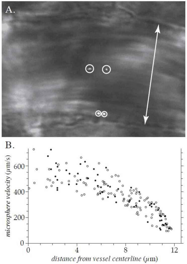
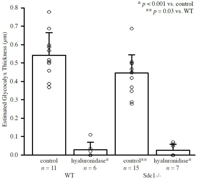
The existence of a hydrodynamically relevant endothelial glycocalyx has been established in capillaries, venules, and arterioles in vivo. The glycocalyx is thought to consist primarily of membrane-bound proteoglycans with glycosaminoglycan side-chains, membrane-bound glypicans, and adsorbed plasma proteins. The proteoglycans found on the luminal surface of endothelial cells are syndecans-1, -2, and -4, and glypican-1. The extent to which any of these proteins might serve to anchor the glycocalyx to the endothelium has not yet been determined. To test whether syndecan-1, in particular, is an essential anchoring protein, we performed experiments to determine the hydrodynamically relevant glycocalyx thickness in syndecan-1 deficient (Sdc1(-/-)) mice. Micro-particle image velocimetry data were collected using a previously described method. Microviscometric analysis of these data consistently revealed the existence of a hydrodynamically relevant endothelial glycocalyx in Sdc1(-/-) mice in vivo. The mean glycocalyx thickness found in Sdc1(-/-) mice was 0.45±0.10 μm (N=15), as compared with 0.54±0.12 μm (N=11) in wild-type (WT) mice (p=0.03). The slightly thinner glycocalyx observed in Sdc1(-/-) mice relative to WT mice may be due to the absence of syndecan-1. These findings show that healthy Sdc1(-/-) mice are able to synthesize and maintain a hydrodynamically relevant glycocalyx, which indicates that syndecan-1 is not an essential anchoring protein for the glycocalyx in Sdc1(-/-) mice. This may also be the case for WT mice; however, Sdc1(-/-) mice might adapt to the lack of syndecan-1 by increasing the expression of other proteoglycans. In any case, syndecan-1 does not appear to be a prerequisite for the existence of an endothelial glycocalyx.


Figures

Figure 1
Figure 1. Typical image and data obtained in vivo
These results are from a 25.1-μm-diameter venule in the cremaster muscle of an Sdc1−/− mouse, prior to hyaluronidase treatment. Shown in (A) is the brightfield image of the vessel, merged with the simultaneously acquired dual images of two fluorescent microspheres (encircled). The double arrow indicates the diameter of the venule. The full μ-PIV data set for this venule, folded onto one-half of the vessel cross-section, is shown in (B). The closed circles indicate data points measured from the vessel wall that is nearer to the top of the image in (A) and open circles indicate data points measured from the opposite wall. The velocity and radial location were measured for 172 microspheres in this venule. The monotonically filtered subset of this data is shown in Figure 2(A).
Figure 2
Figure 2. Representative results of μ-PIV and microviscometric analysis in venules of Sdc1−/− mice in vivo
Shown in the left column are results from a 25.1-μm-diameter venule before hyaluronidase treatment. In the right column are results from a 48.2-μm-diameter venule after hyaluronidase treatment to degrade the glycocalyx. Panels A and B show the monotonically filtered μ-PIV data (circles) and the fitted axisymmetric velocity profile (curve) corresponding to trial glycocalyx thickness that produced the smallest least-squares error of all admissible trial thicknesses. The shaded region near the vessel wall represents the glycocalyx. The corresponding relative blood viscosity (solid curve) and shear-rate (dashed curve) distributions are shown in panels C and D. Panels E and F show the variation in the normalized least-squares error associated with the fit to the μ-PIV data. The estimated glycocalyx thickness is 0.42 μm in the untreated venule and 0.0 μm in the hyaluronidase-treated venule.
Figure 3
Figure 3. Representative results of μ-PIV and microviscometric analysis in venules of WT mice in vivo, with the same interpretation as Figure 2
Shown in the left column are results from a 43.7-μm-diameter venule before hyaluronidase treatment. In the right column are results from a 55.3-μm-diameter venule after hyaluronidase treatment to degrade the glycocalyx. The estimated hydrodynamically relevant glycocalyx thickness is 0.50 μm in the untreated venule and 0.02 μm in the hyaluronidase-treated venule.
Figure 4
Figure 4. Mean estimated glycocalyx thickness in venules of Sdc1−/− and WT mice before and after hyaluronidase treatment
The bars show the mean and standard deviation for each group. The open circles indicate the estimated glycocalyx thickness for each individual vessel in the data set. In the untreated Sdc1−/− group, the mean glycocalyx thickness was found to be 0.45±0.10 μm (n=15 venules, N=6 mice). This is ~15% thinner than the 0.54±0.12 μm glycocalyx found in WT mice (n=11, N=7, p=0.03). After hyaluronidase treatment, the mean glycocalyx thickness was 0.03±0.03 μm in Sdc1−/− mice (n=7, N=3, p<0.001 vs. Sdc1−/− or WT mice before treatment) and 0.03±0.04 μm in WT mice (n=6, N=3, p<0.001 vs. Sdc1−/− or WT mice before treatment).
Figure 5
Figure 5. Leukocyte Adhesion in Sdc1−/− and WT mice. This chart shows the percentage of vessel segments in each group having 0, 1, 2, 3 or 4 adherent leukocytes
None of the vessels observed had greater than 4 adherent leukocytes visible in the mid-sagittal plane. In Sdc1−/− mice, 42% of vessels studied had 2 or more adherent leukocytes, whereas in WT mice this number dropped to 11%. The mean number of adherent leukocytes was 1.5 ±1.3 (n=12) in Sdc1−/− mice and 0.7±0.8 (n=18; p=0.04) in WT mice.

References

    1. Alexander CM, Reichsman F, Hinkes MT, Lincecum J, Becker KA, Cumberledge S, Bernfield M. Syndecan-1 is required for Wnt-1-induced mammary tumorigenesis in mice. Nat. Gen. 2000;25:329–332. - PubMed
    1. Alexopoulou AN, Multhaupt HAB, Couchman JR. Syndecans in wound healing, inflammation, and vascular biology. Int. J. Biochem. Cell Biol. 2007;39:505–528. - PubMed
    1. Baez S. An open cremaster muscle preparation for the study of blood vessels by in vivo microscopy. Microvasc. Res. 1973;5:384–394. - PubMed
    1. Barker AL, Konopatskaya O, Neal CR, Macpherson JV, Whatmore JL, Winlove PC, Unwin PR, Shore AC. Observation and characterisation of the glycocalyx of viable human endothelial cells using confocal laser scanning microscopy. Int. Phys. Chem. Chem. Phys. 2004;6:1006–1011.
    1. Bernfield M, Kokenyesi R, Kato M, Hinkes MT, Spring J, Gallo RL, Lose EJ. Biology of the syndecans: a family of transmembrane heparan-sulfate proteoglycans. Annu. Rev. Cell Biol. 1992;8:365–393. - PubMed

Publication types

LinkOut - more resources