

Mediastinal leiomyosarcoma concurrent with intra-aortic thrombosis
- PMID: 23429014
- PMCID: PMC3603919
- DOI: 10.1136/bcr-2012-007527
Mediastinal leiomyosarcoma concurrent with intra-aortic thrombosis
Abstract
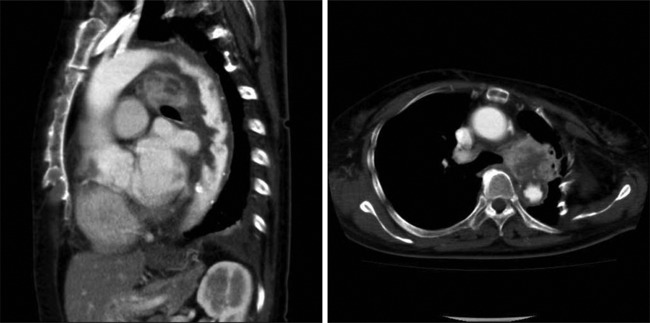
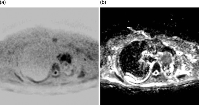
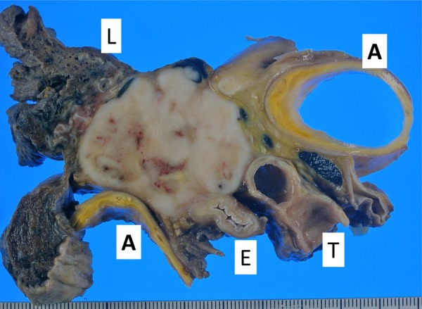
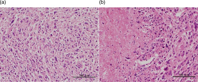
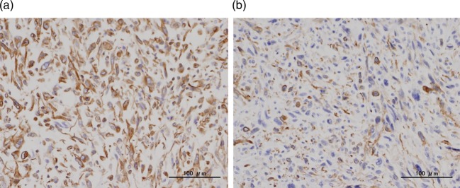
We report a case of a large intra-aortic thrombosis in an 83-year-old woman concurrent with metastatic mediastinal leiomyosarcoma. Imaging studies incidentally detected a mediastinal malignant tumour metastasising to bilateral adrenals and an extensive intra-aortic mass that was suspected to be intra-aortic thrombosis. One month later massive embolism developed in the lower limb and her condition deteriorated rapidly resulting in death. Autopsy revealed diffused proliferation of highly pleomorphic atypical cells accompanied by necrosis in the mediastinum tumours and bilateral adrenal glands. Leiomyosarcoma metastasising to bilateral adrenals was confirmed by the results of immunostaining. The intra-aortic mass suggested that the fragmented thrombus might be the cause of a sudden lower-limb embolism. Microscopic examination showed that the mass lesion in the aortic arch was composed of a blood clot containing neutrophils. We report this case because leiomyosarcoma arising from the mediastinum and, especially, associated with an extraordinarily large intra-aortic thrombosis is very rare.
Figures
References
-
- Tsilimparis N, Hanack U, Pisimisis G, et al. Thrombus in the non-aneurysmal, non-atherosclerotic descending thoracic aorta—an unusual source of arterial embolism. Eur J Vasc Endovasc Surg 2011;41:450–7 - PubMed
-
- Abiko T, Sato S, Futamata T, et al. A case of pleomorphic leiomyosarcoma of the posterior mediastinum. J Japanese Assoc Chest Surg 2005;19:819–22
-
- Farshid G, Pradhan M, Goldblum J, et al. Leiomyosarcoma of somatic soft tissues: a tumor of vascular origin with multivariate analysis of outcome in 42 cases. Am J Surg Pathol 2002;26:14–24 - PubMed
-
- Reber PU, Patel AG, Stauffer E, et al. Mural aortic thrombi: An important cause of peripheral embolization. J Vasc Surg 1999;30:1084–9 - PubMed
Publication types
MeSH terms
LinkOut - more resources
Full Text Sources
Other Literature Sources
Medical