

The role of lipid domains in bacterial cell processes
- PMID: 23429192
- PMCID: PMC3588084
- DOI: 10.3390/ijms14024050
The role of lipid domains in bacterial cell processes
Abstract
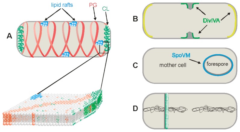
Membranes are vital structures for cellular life forms. As thin, hydrophobic films, they provide a physical barrier separating the aqueous cytoplasm from the outside world or from the interiors of other cellular compartments. They maintain a selective permeability for the import and export of water-soluble compounds, enabling the living cell to maintain a stable chemical environment for biological processes. Cell membranes are primarily composed of two crucial substances, lipids and proteins. Bacterial membranes can sense environmental changes or communication signals from other cells and they support different cell processes, including cell division, differentiation, protein secretion and supplementary protein functions. The original fluid mosaic model of membrane structure has been recently revised because it has become apparent that domains of different lipid composition are present in both eukaryotic and prokaryotic cell membranes. In this review, we summarize different aspects of phospholipid domain formation in bacterial membranes, mainly in Gram-negative Escherichia coli and Gram-positive Bacillus subtilis. We describe the role of these lipid domains in membrane dynamics and the localization of specific proteins and protein complexes in relation to the regulation of cellular function.
Figures
References
-
- Govindarajan S., Nevo-Dinur K., Amster-Choder O. Compartmentalization and spatiotemporal organization of macromolecules in bacteria. FEMS Microbiol. Rev. 2012;36:1005–1022. - PubMed
-
- Matsumoto K., Kusaka J., Nishibori A., Hara H. Lipid domains in bacterial membranes. Mol. Microbiol. 2006;61:1110–1117. - PubMed
-
- Singer S.J., Nicolson G.L. The fluid mosaic model of the structure of cell membranes. Science. 1972;175:720–731. - PubMed
LinkOut - more resources
Full Text Sources
Other Literature Sources
Research Materials
