

Ectopic adrenal cortical adenoma in the gastric wall: case report
- PMID: 23429246
- PMCID: PMC3574608
- DOI: 10.3748/wjg.v19.i5.778
Ectopic adrenal cortical adenoma in the gastric wall: case report
Abstract
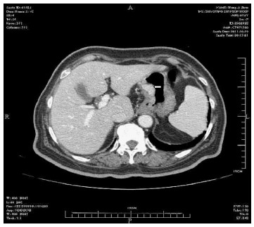
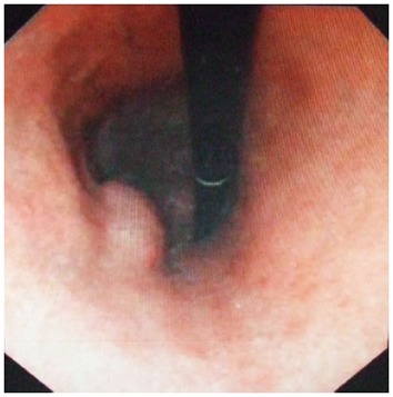
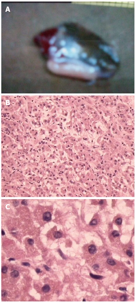
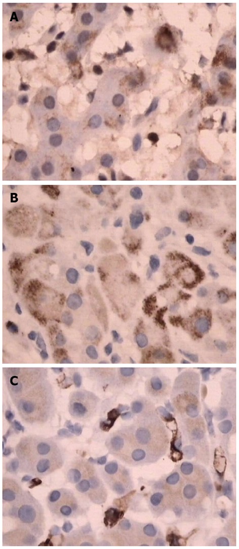
Ectopic adrenal cortical neoplasms are extremely rare. Ectopic adrenocortical tissue can be found in locations such as the celiac axis, the broad ligament, the adnexa of the testes, and the spermatic cord; however, they rarely involve the stomach. We report an unusual case of a patient with an ectopic adrenal cortical adenoma in the gastric wall. The patient was a 72-year old female admitted to our hospital with upper abdominal discomfort. Physical examination revealed tenderness below the xiphoid process. Both computed tomography and fibergastroscopy revealed a mass on the lesser curvature side of the gastric antrum; it was initially diagnosed as a gastric stromal tumor. After adequate preparation, the patient underwent surgery. During the procedure, we found a 30 mm × 30 mm mass with medium density in the lesser curvature near the gastric antrum within the serosa. Following immunohistochemistry examination, we corrected the diagnosis to an ectopic adrenal cortical adenoma; the tumor was nonfunctional.
Keywords: Adrenal adenoma; Adult; Ectopic adrenal cortical neoplasms; Nonfunctional adenoma; Stomach.
Figures
References
-
- Barwick TD, Malhotra A, Webb JA, Savage MO, Reznek RH. Embryology of the adrenal glands and its relevance to diagnostic imaging. Clin Radiol. 2005;60:953–959. - PubMed
-
- Graham LS. Celiac accessory adrenal glands. Cancer. 1953;6:149–152.
Publication types
MeSH terms
Substances
LinkOut - more resources
Full Text Sources
Other Literature Sources
Medical
