. 2013 Jun;20(6):785-99.
doi: 10.1038/cdd.2013.10. Epub 2013 Feb 22.

Intracellular localization of the BCL-2 family member BOK and functional implications

Affiliations

Intracellular localization of the BCL-2 family member BOK and functional implications

N Echeverry et al. Cell Death Differ. 2013 Jun.

Abstract

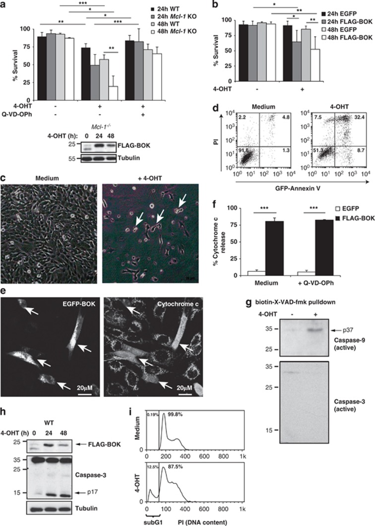
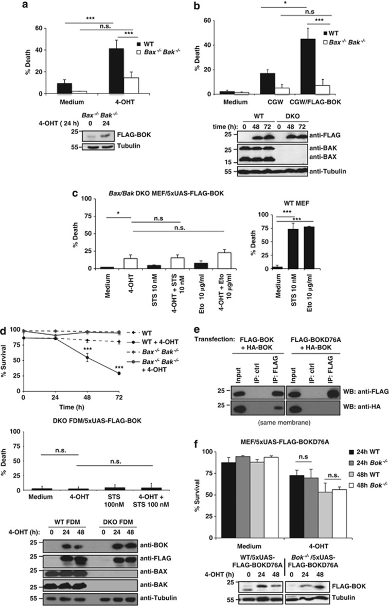
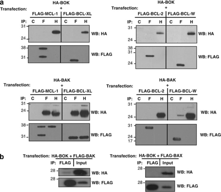
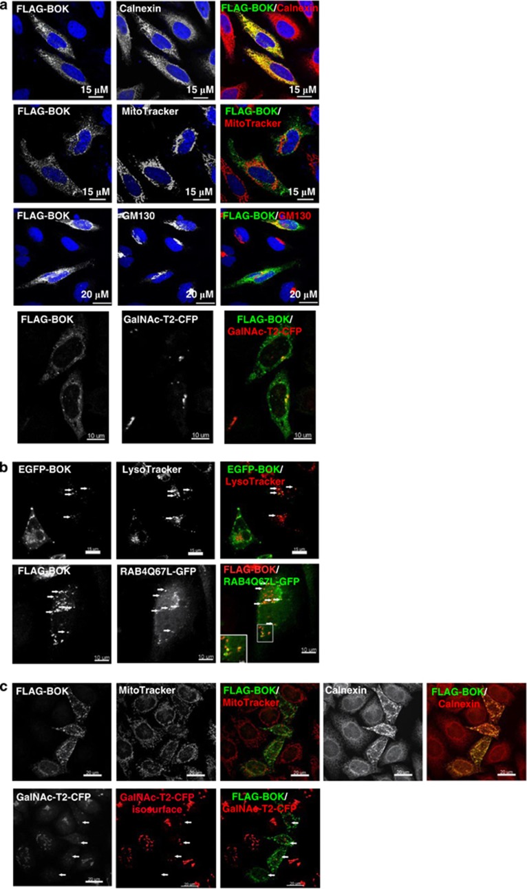
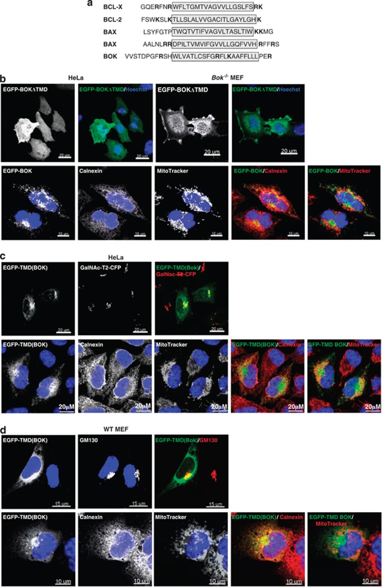
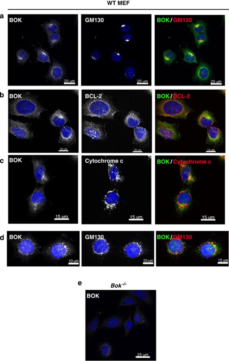
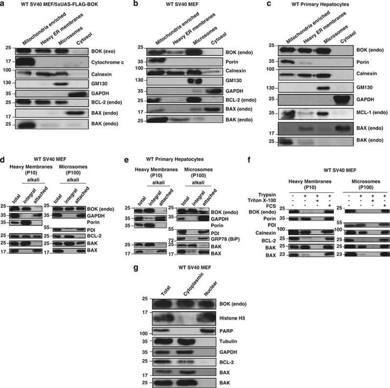
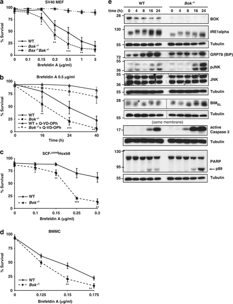
The pro-apoptotic BCL-2 family member BOK is widely expressed and resembles the multi-BH domain proteins BAX and BAK based on its amino acid sequence. The genomic region encoding BOK was reported to be frequently deleted in human cancer and it has therefore been hypothesized that BOK functions as a tumor suppressor. However, little is known about the molecular functions of BOK. We show that enforced expression of BOK activates the intrinsic (mitochondrial) apoptotic pathway in BAX/BAK-proficient cells but fails to kill cells lacking both BAX and BAK or sensitize them to cytotoxic insults. Interestingly, major portions of endogenous BOK are localized to and partially inserted into the membranes of the Golgi apparatus as well as the endoplasmic reticulum (ER) and associated membranes. The C-terminal transmembrane domain of BOK thereby constitutes a 'tail-anchor' specific for targeting to the Golgi and ER. Overexpression of full-length BOK causes early fragmentation of ER and Golgi compartments. A role for BOK on the Golgi apparatus and the ER is supported by an abnormal response of Bok-deficient cells to the Golgi/ER stressor brefeldin A. Based on these results, we propose that major functions of BOK are exerted at the Golgi and ER membranes and that BOK induces apoptosis in a manner dependent on BAX and BAK.


Figures

Figure 1
Figure 1
Overexpressed BOK activates the intrinsic apoptotic pathway in SV40-immortalized MEF. (a) SV40-immortalized WT and Mcl-1−/− MEF/5xUAS-FLAG-BOK cells were treated as indicated with 4-OHT (0.1 μM) and Q-VD-OPh (25 μM) for 24 or 48 h and viability assessed by GFP-Annexin V/PI staining and flow cytometry. Data are presented as means ±S.D. from three independent experiments. Induction of FLAG-BOK in SV40-immortalized WT (see h) and Mcl-1−/− MEF was controlled by Western blotting. (b) SV40-immortalized WT MEF/5xUAS-FLAG-BOK and WT MEF/5xUAS-EGFP cells were treated with 4-OHT (0.1 μM) for 24 or 48 h and viability assessed by PI staining and flow cytometry. Data represent means±S.D. from 6 (FLAG-BOK) and 5 (EGFP) independent experiments, respectively. (c) Wide-field micrographs of WT MEF/5xUAS-FLAG-BOK cells induced for 48 h with 0.1 μM 4-OHT. White arrows indicate apoptotic cells characterized by plasma membrane blebbing, rounding and detachment. (d) Representative dot plots (n≥6) of GFP–Annexin V/PI staining of MEF/5xUAS-FLAG-BOK cells with or without FLAG-BOK induction for 48 h. (e) Confocal micrographs of WT MEF transiently transfected with EGFP-BOK for 9 h and immunostained with an antibody against cytochrome c. (f) WT MEF were transfected with expression constructs for EGFP or FLAG-BOK, in the absence or presence of 20 μM Q-VD-OPh, for 9 h and percentages of EGFP- or FLAG-BOK-positive cells with released cytochrome c determined by counting 100 cells for each condition. Data represent means ± S.D. of three independent experiments. (g) WT MEF/5xUAS-FLAG-BOK cells were left untreated or treated with 0.1 μM 4-OHT for 14 h. Active caspases were covalently labeled with biotinylated X-VAD-fmk, pulled down with streptavidin–sepharose beads and Western blots probed for caspase-9 and caspase-3. Whereas active caspase-9 was pulled down, no signal for active caspase-3 could be detected at this specific time point. (h) WT MEF/5xUAS-FLAG-BOK cells were treated with 4-OHT (0.1 μM) for the indicated times and total protein lysates analyzed by Western blotting using anti-BOK and anti-caspase-3 antibodies. (i) Representative histograms (n⩾3) showing DNA content of WT MEF/5xUAS-FLAG-BOK with or without FLAG-BOK induction (t=48 h). The cell population with fragmented genomic DNA is indicated as subG1. Significance values were determined by paired Student's t-test (***P<0.005; **P<0.01; *P<0.05)
Figure 2
Figure 2
BOK-induced apoptosis is largely BAX/BAK-dependent. (a) FLAG-BOK was induced in SV40-immortalized WT and Bax−/−Bak−/− MEF/5xUAS-FLAG-BOK cells by addition of 0.1 μM 4-OHT. Cell death was determined after 48 h by flow cytometric analysis (after staining with GFP-Annexin V and PI). Data are presented as means ± S.D. from greater than three independent experiments. (b) WT and Bax−/−Bak−/− SV40 MEF were transduced with CAD-G-Whiz empty vector (CGW) or CGW/FLAG-BOK for 40 h and apoptosis of EGFP-positive cells measured by flow cytometry (Cherry-Annexin V-positive cells). Data are presented as means±S.D. from three independent experiments. FLAG-BOK expression and correct genotypes were verified by Western blotting, using antibodies to BOK, BAX and BAK. (c) FLAG-BOK was induced in Bax−/−Bak−/− MEF/5xUAS-FLAG-BOK cells for 24 h, followed by treatment with 10 nM staurosporine or 10 μg/ml etoposide for an additional 24 h. Cell death was quantified as described in (a). WT SV40 MEF treated with the same concentration of drugs for 24 h are shown as a control. Data represent means ± S.D. from three independent experiments. (d) Survival of WT and Bax−/−Bak/ FDM/5xUAS-FLAG-BOK cells with or without induction of FLAG-BOK expression (upper panel). Surviving cells were determined by flow cytometry and data represent GFP-Annexin V/PI double-negative cells. Data represent means ± S.D. from three independent experiments. Lower panel: FLAG-BOK was induced for 8 h in Bax−/−Bak/ FDM/5xUAS-FLAG-BOK cells, followed by treatment with 100 nM staurosporine for an additional 24 h. Data represent means ± S.D. from greater than three independent experiments. FLAG-BOK expression and correct genotypes were verified by Western blotting, using antibodies to FLAG, BOK (RabMab BOK-1-5), BAX and BAK. (e) Co-immunoprecipitation assay in HEK 293T cells transiently co-transfected for 14 h with HA-BOK/pcDNA3 and FLAG-BOK/pcDNA3 or FLAG-BOK(D76A)/pcDNA3. Anti-FLAG and non-specific mouse IgG1 (isotype control) immunoprecipitates were probed by Western blotting using HA- and FLAG-specific antibodies. (f) Viability of WT and Bok−/− SV40 MEF upon induction of FLAG-BOK(D76A) was determined by GFP-Annexin V/PI negativity by flow cytometry. Induction of FLAG-BOK(D76A) was controlled by Western blotting using anti-BOK antibody. Data are presented as means±S.D. of greater than three independent experiments. Significance was determined by Student's t-test (***P<0.005, **P<0.01 and *P<0.05; n.s.: not significant)
Figure 3
Figure 3
BOK does not co-immunoprecipitate with other BCL-2 family members. (a) HA-BOK or HA-BAK were transiently co-expressed with FLAG-tagged MCL-1, BCL-XL, BCL-2 or BCL-W in HEK 293T cells. Immunoprecipitates (C: protein G sepharose beads only; F: anti-FLAG; H: anti-HA) were probed by Western blotting with anti-FLAG and anti-HA antibodies. (b) HA-BOK was transiently co-transfected with FLAG-BAX or FLAG-BAK. Anti-FLAG immunoprecipitates were probed by Western blot using anti-HA and anti-FLAG antibodies
Figure 4
Figure 4
Overexpressed BOK mainly localizes to membranes of the endoplasmic reticulum, Golgi apparatus and associated membranes. (a) Representative confocal scanning micrographs of HeLa cells transiently transfected with FLAG-BOK/pcDNA3 for 18 h and stained with organelle-specific antibodies against calnexin (endoplasmic reticulum) or GM130 (Golgi matrix), MitoTracker Deep Red 633 (mitochondria), GalNAc-T2-CFP (trans and medial Golgi) and Hoechst 33342 (nuclei). (b) HeLa cells were transfected with pEGFP-BOK for 8 h. Images were taken at real time. EGFP-BOK is found in vesicular patterns and partially colocalizes with the acidic organelle marker LysoTracker, as indicated by white arrows. FLAG-BOK and GFP-RAB4Q67L-GFP (constitutive active mutant, endosomal marker) were co-expressed in HeLa cells for 22 h. Partial colocalization is indicated by white arrows and in the magnified inlet. (c) HeLa cells stably expressing GaLNAc-T2-CFP (specific marker for trans and medial Golgi) were transfected with FLAG-BOK/pcDNA3 for 21 h in the presence of 25 μM Q-VD-OPh. Cells were stained with antibodies to calnexin and MitoTracker Deep Red 633. Forty Z-stacks were acquired and GaLNaC-T2-CFP channel iso-surface 3D Golgi reconstruction was performed using the IMARIS software. Cells expressing FLAG-BOK (white arrows) show weakened GalNAc-T2-CFP signal intensity, correlating with increased Golgi dissociation and fragmentation compared with non-transfected cells
Figure 5
Figure 5
The transmembrane domain of BOK is necessary and sufficient for membrane targeting. (a) Amino-acid sequences of the C-terminal transmembrane domains (TMD) of BOK, BCL-2, BCL-X, BAX and BAK. Positively charged amino acids are printed in bold. (b) Confocal scanning micrographs of HeLa cells and SV40-immortalized Bok−/− MEF transfected with pEGFP-BOKΔTMD for 18 and 8 h, respectively, and stained as indicated. pEGFP-BOKΔTMD shows cytosolic and nuclear localization (Hoechst 33342, blue). EGFP-(WT)BOK is shown as control. (c) HeLa and HeLa/GalNAc-T2-CFP cells were transfected with pEGFP-TMD(BOK) for 24 h and stained with antibodies to calnexin (ER), MitoTracker Deep Red 633 (mitochondria) and Hoechst 33342 (DNA). EGFP-TMD(BOK) localizes extensively to Golgi membranes and ER membranes. (d) Confocal micrographs of SV40-immortalized WT MEF transiently transfected with GFP-TMD(BOK) for 24 h and stained with the indicated organelle markers
Figure 6
Figure 6
Endogenous BOK strongly associates with the Golgi and ER compartments. (a–c) Confocal scanning micrographs of SV40-immortalized WT MEF stained with Hoechst 33342 (nuclei) and antibodies against BOK, GM130 (cis-Golgi marker), BCL-2 and cytochrome c. (d) WT SV40 MEF were treated with 0.3 μg/ml brefeldin A for 15 h and stained with anti-BOK and anti-GM130 antibodies and Hoechst 33342. (e) SV40-immortalized Bok−/− MEF were stained with antibodies to BOK and Hoechst 33342
Figure 7
Figure 7
Analysis of the localization of overexpressed and endogenous BOK by subcellular fractionation and membrane insertion assays. Anti-BOK Western blots of subcellular fractions prepared from SV40-immortalized WT MEF/5xUAS-FLAG-BOK cells after FLAG-BOK induction with 0.1 μM 4-OHT for 14 h (a), SV40 WT MEF (b) and primary hepatocytes freshly isolated from young adult C57BL/6 (WT) mice (c). Membranes were reprobed with antibodies to mouse BCL-2, MCL-1, BAX and BAK for comparison. Reprobing of the membranes with antibodies to Porin or cytochrome c (mitochondria), calnexin (ER), GM130 (Golgi) and GAPDH (cytosol) served as controls for the purity of the subcellular fractions. Heavy P10 and light P100 membrane fractions from SV40 WT MEF (d) and mouse primary hepatocytes (e) were extracted directly with detergent (total) or first treated with sodium carbonate solution (pH 12) to remove loosely attached membrane proteins. Remaining membrane proteins were lysed in detergent and all fractions analyzed by Western blotting for endogenous BOK and indicated proteins that served as controls for integrally inserted membrane proteins (Porin, BAK, BCL-2), or loosely attached and soluble proteins (BAX, GAPDH). BiP and PDI are luminal ER proteins and probing for them served as a control for vesicle integrity. (f) Heavy and light membranes were prepared from SV40 WT MEF and membrane proteins facing the cytosol, including endogenous BOK, were digested with 0.01% trypsin. (g) Purified nuclear and cytoplasmic fractions were prepared from SV40 WT MEF. A large portion of endogenous BOK was found in the purified nuclear fraction. The purity of the fractions was controlled by reprobing the membrane with antibodies against Histone H3 and PARP (nuclear) as well as Tubulin and GAPDH (cytoplasmic). In contrast to BOK, no endogenous BCL-2, BAX or BAK could be detected in the nuclear fraction
Figure 8
Figure 8
BOK-deficient cells show an atypical response to treatment with brefeldin A. (a) The survival of SV40-immortalized WT, Bok−/− and Bax−/−Bak−/− MEF treated with the indicated doses of brefeldin A for 24 h was quantified by flow cytometry (GFP-Annexin V/PI negative cells). (b) Time kinetics of cell survival of SV40 WT and Bok−/− MEF treated with 0.5 μg/ml brefeldin A with or without 25 μM Q-VD-OPh. (c) WT and Bok−/− SCF-condHoxb8-immortalized myeloid progenitors were treated with the indicated doses of brefeldin A for 18 h. Cell survival was determined by GFP-Annexin V/PI staining followed by flow cytometric analysis. (d) Brefeldin A dose-response curves (treatment for 24 h) of ex vivo generated bone marrow-derived mast cells (BMMC) derived from WT or Bok−/− mice. (e) Western blot analysis of protein lysates prepared from SV40 WT and Bok−/− MEF that had been treated with 3 μg/ml brefeldin A for the indicated times. Data are representative of three independent experiments using different SV40-immortalized MEF lines. All quantitative data (dose responses and time kinetics) are presented as means±S.D. of at least five (MEF) or three (BMMC, SCF-condHoxb8) independent experiments, respectively. Three independent lines (each derived from a different embryo) were used for SV40-immortalized WT and Bok−/− MEF, respectively. Significance was determined by unpaired Student's t-test (***P<0.005, **P<0.01 and *P<0.05)

References

    1. Youle RJ, Strasser A. The BCL-2 protein family: opposing activities that mediate cell death. Nat Rev Mol Cell Biol. 2008;9:47–59. - PubMed
    1. Schinzel A, Kaufmann T, Borner C. Bcl-2 family members: integrators of survival and death signals in physiology and pathology [corrected] Biochim Biophys Acta. 2004;1644:95–105. - PubMed
    1. Adams JM, Cory S. Life-or-death decisions by the Bcl-2 protein family. Trends Biochem Sci. 2001;26:61–66. - PubMed
    1. Giam M, Huang DC, Bouillet P. BH3-only proteins and their roles in programmed cell death. Oncogene. 2008;27 (Suppl 1:S128–S136. - PubMed
    1. Chipuk JE, Moldoveanu T, Llambi F, Parsons MJ, Green DR. The BCL-2 family reunion. Mol Cell. 2010;37:299–310. - PMC - PubMed

Publication types

MeSH terms

Substances