. 2013 Jul;62(1):26-40.
doi: 10.1097/FJC.0b013e31828bc88a.

Subtype-specific estrogen receptor-mediated vasodilator activity in the cephalic, thoracic, and abdominal vasculature of female rat

Affiliations

Subtype-specific estrogen receptor-mediated vasodilator activity in the cephalic, thoracic, and abdominal vasculature of female rat

Ossama M Reslan et al. J Cardiovasc Pharmacol. 2013 Jul.

Abstract

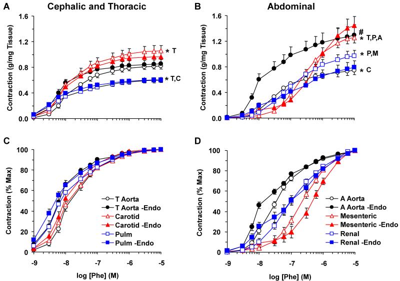
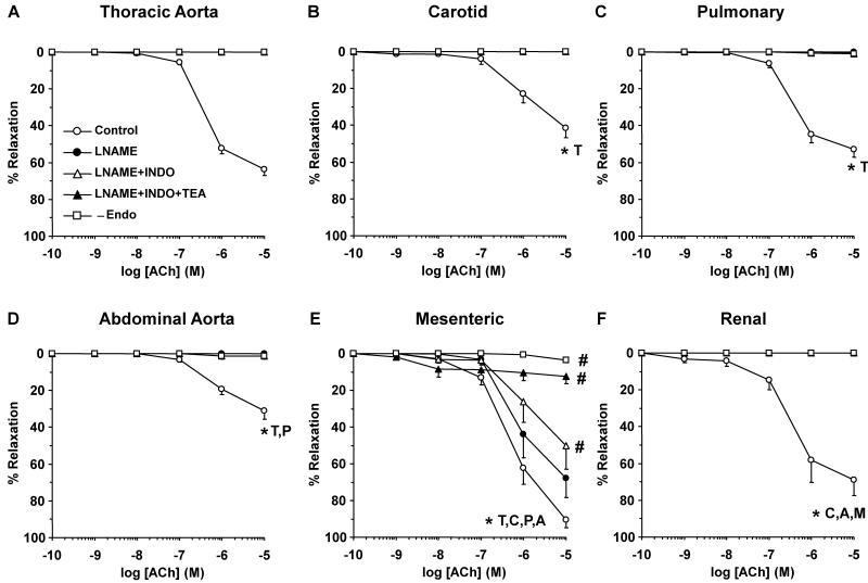
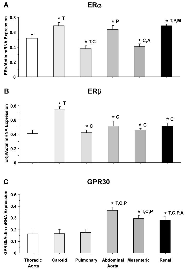
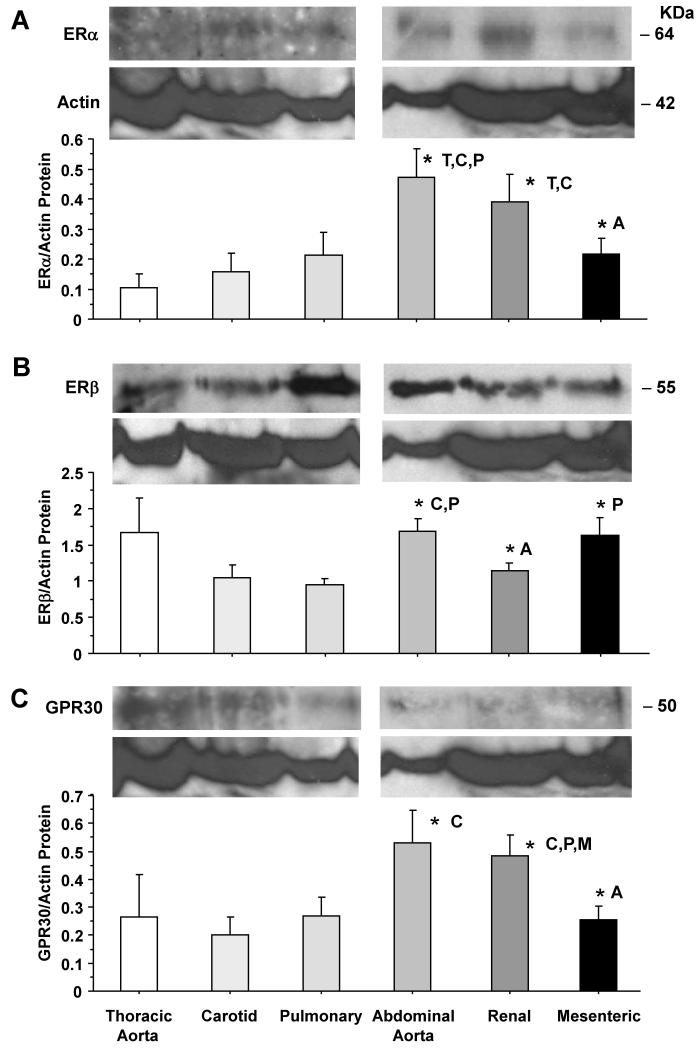
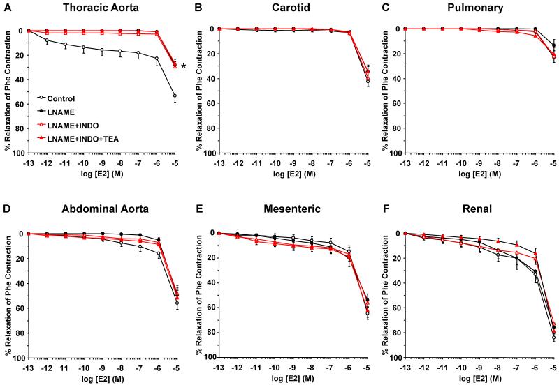
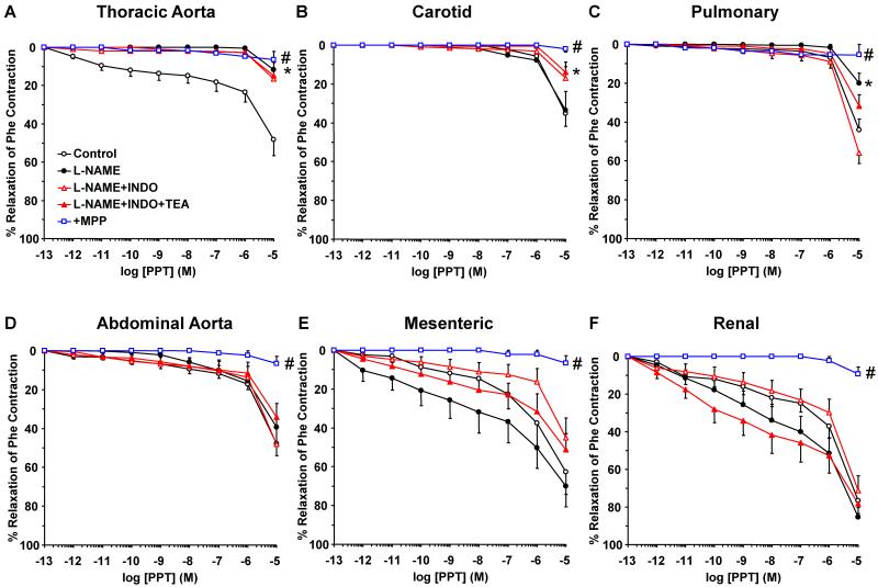
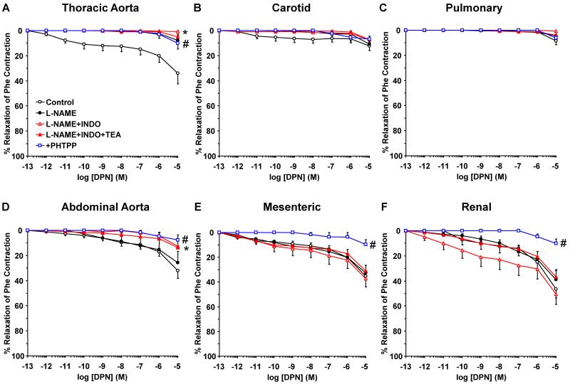
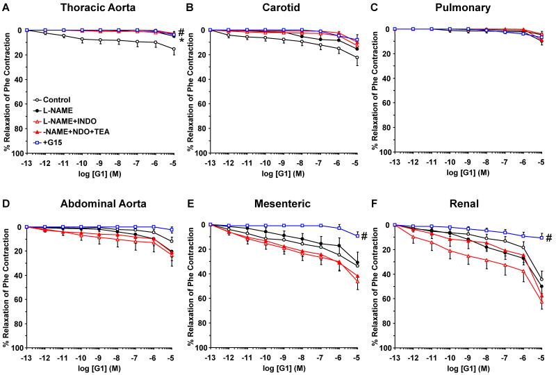
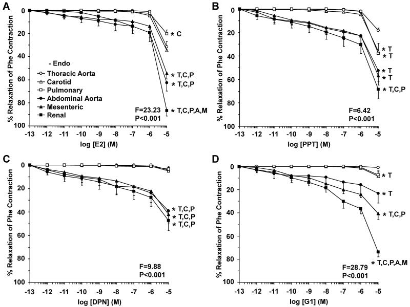
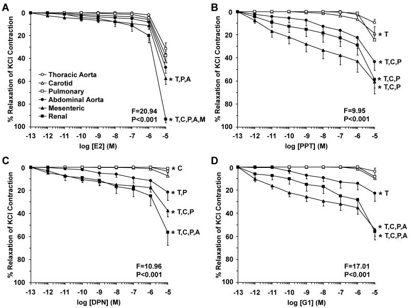
Estrogen receptors (ERs) mediate genomic and nongenomic vasodilator effects, but estrogen therapy may not provide systemic vascular protection. To test whether this is because of regional differences in ER distribution or vasodilator activity, cephalic (carotid artery), thoracic (thoracic aorta and pulmonary artery), and abdominal arteries (abdominal aorta, mesenteric artery, and renal artery) from female Sprague-Dawley rats were prepared to measure contraction to phenylephrine and relaxation to acetylcholine (ACh) and the ER activators 17β-estradiol (E2) (all ERs), 4,4',4″-(4-propyl-[1H]-pyrazole-1,3,5-triyl)-tris-phenol (PPT) (ERα), diarylpropionitrile (DPN) (ERβ), and (±)-1-[(3aR*,4S*,9bS*)-4-(6-bromo-1,3-benzodioxol-5-yl)-3a,4,5,9b-tetrahydro-3H-cyclopenta[c]quinolin-8-yl]-ethanone (G1) (GPR30). Phenylephrine caused contraction that was enhanced in endothelium-denuded aorta, supporting endothelial release of vasodilators. In cephalic and thoracic arteries, ACh relaxation was abolished by the nitric oxide (NO) synthase inhibitor Nω-nitro-L-arginine methyl ester (L-NAME), suggesting a role of NO. In mesenteric vessels, ACh-induced relaxation was partly inhibited by the L-NAME + cyclooxygenase inhibitor indomethacin and blocked by the K+ channel blocker tetraethylammonium, suggesting a hyperpolarization pathway. E2 and PPT caused similar relaxation in all vessels. DPN and G1 caused smaller relaxation that was more prominent in abdominal vessels. Reverse transcription-polymerase chain reaction revealed variable ERα messenger RNA expression and increased ERβ in carotid artery and GPR30 in abdominal arteries. Western blots revealed greater amounts of ERα, ERβ, and GPR30 in abdominal arteries. In thoracic aorta, E2-, PPT-, and DPN-induced relaxation was blocked by L-NAME and was associated with increased nitrite/nitrate production, suggesting a role of NO. In abdominal vessels, E2-, PPT-, DPN-, and G1-induced relaxation persisted in L-NAME + indomethacin + tetraethylammonium-treated or endothelium-denuded arteries, suggesting direct effect on vascular smooth muscle. E2, PPT, DPN, and G1 caused greater relaxation of KCl-induced contraction in abdominal vessels, suggesting inhibitory effects on Ca2+ entry. Thus, E2 and ERα stimulation produces similar relaxation of the cephalic, thoracic, and abdominal arteries. In the cephalic and thoracic arteries, particularly the thoracic aorta, E2-induced and ERα- and ERβ-mediated vasodilation involves NO production. ERβ- and GPR30-mediated relaxation is greater in the abdominal arteries and seems to involve hyperpolarization and inhibition of vascular smooth muscle Ca2+ entry. Specific ER agonists could produce vasodilation in specific vascular beds without affecting other vessels in the systemic circulation.


Figures

Fig. 1
Fig. 1
Phe-induced contraction in cephalic, thoracic and abdominal arteries of female rat. Endothelium-intact and -denuded segments of thoracic aorta, carotid and pulmonary artery (A) as well as abdominal aorta, mesenteric and renal artery (B) were incubated in normal Krebs solution. The vessels were stimulated with the α-adrenergic agonist Phe (10−9 to 10−5 M) and the contractile response was recorded and presented in g/mg tissue (Upper Panels) or as % of maximal contraction (Lower Panels). The Phe response in intact vessels (+Endo) was compared with the response in endothelium-denuded vessels (-Endo). Data represent means±SEM, n=8 to 10. * Significantly different maximal contraction (p<0.05).
Fig. 2
Fig. 2
Acetylcholine (ACh)-induced relaxation in cephalic, thoracic and abdominal vessels of female rat. Endothelium-intact and -denuded segments of thoracic aorta (A), carotid (B) and pulmonary artery (C) as well as abdominal aorta (D), mesenteric (E) and renal artery (F) were precontracted with submaximal concentration of Phe (10−6 M in mesenteric and renal artery, 3×10⁻⁷ M in the other arteries) and the steady-state contraction was recorded. ACh (10−9 to 10−5 M) was added and the % relaxation of Phe contraction was compared in blood vessels non-treated or treated with the NOS inhibitor L-NAME (3×10⁻⁴ M), COX inhibitor indomethacin (INDO, 10−5 M), and BKCa blocker TEA (30 mM). ACh-induced relaxation was also compared in intact versus endothelium-denuded arteries (-Endo). Data represent means±SEM, n=8 to 10. * Significantly different maximal relaxation (p<0.05).
Fig. 3
Fig. 3
ER-mediated relaxation in cephalic, thoracic and abdominal arteries of female rat. Endothelium-intact segments of thoracic aorta (open circles), carotid (open triangles) and pulmonary artery (open squares) as well as abdominal aorta (closed circles), mesenteric (closed triangles) and renal artery (closed squares) were precontracted with submaximal concentration of Phe and the steady-state contraction was recorded. Increasing concentrations (10−12 to 10−5 M) of 17β-estradiol (E2, activator of most ERs) (A), PPT (ERα agonist) (B), DPN (ERβ agonist) (C), or G1 (GPR30 agonist) (D) were added and the % relaxation of Phe contraction was measured. Data represent means±SEM, n= 8 to 10. * Significantly different (p<0.05) from corresponding measurement in thoracic aorta [T], carotid [C], pulmonary [P], abdominal aorta [A], mesenteric [M] and renal artery [R].
Fig. 4
Fig. 4
ERα, ERβ and GPR30 mRNA expression in cephalic, thoracic and abdominal arteries of female rat. Tissue homogenate of thoracic aorta, carotid, pulmonary, abdominal aorta, mesenteric and renal artery were prepared for real time RT-PCR. The mRNA expression of ERα (A), ERβ (B), and GPR30 (C) was measured and normalized to the house-keeping gene actin. Data represent means±SEM, n=8 to 10. * Significantly different (p<0.05) from corresponding measurement in thoracic aorta [T], carotid [C], pulmonary [P], abdominal aorta [A], mesenteric [M] and renal artery [R].
Fig. 5
Fig. 5
ERα, ERβ and GPR30 protein amount in cephalic, thoracic and abdominal arteries of female rat. Tissue homogenate of thoracic aorta, carotid, pulmonary, abdominal aorta, renal and mesenteric artery were prepared for Western blot analysis. ER subtypes were detected using antibodies to ERα (1:500) (A), ERβ (1:1000) (B) and GPR30 (1:500) (C). Blots for cephalic and thoracic arteries (aorta, carotid, pulmonary) as compared to abdominal arteries (abdominal aorta, renal, mesenteric) were performed on different gels. The representative immunoblots for the abdominal aorta, renal, and mesenteric artery in panels B and C have the same actin because the blots were first reacted with GPR30 antibody, stripped, then reacted with ERβ antibody, stripped, then reacted with actin antibody. The intensity of the immunoreactive bands was analyzed using optical densitometry, and normalized to the house keeping protein actin. Data represent means±SEM, n= 8 to 10. * Significantly different (p<0.05) from corresponding measurement in thoracic aorta [T], carotid [C], pulmonary [P], abdominal aorta [A], mesenteric [M] and renal artery [R].
Fig. 6
Fig. 6
Contribution of NO, PGI2, and hyperpolarization factor to E2-induced relaxation of cephalic, thoracic and abdominal arteries of female rat. Endothelium-intact segments of thoracic aorta (A), carotid (B) pulmonary (C) abdominal aorta (D), mesenteric (E) and renal artery (F) were either nontreated or pretreated for 15 min with L-NAME (3×10⁻⁴ M), L-NAME+indomethacin (INDO, 10−5 M), or L-NAME+INDO+TEA (30 mM). The vessels were precontracted with a submaximal concentration of Phe then increasing concentrations (10−12 to 10−5 M) of 17β-estradiol (E2, activator of most ERs) were added and the % relaxation of Phe contraction was measured. Data represent means±SEM, n= 8 to 10. * Significantly different (p<0.05) from corresponding measurements in control nontreated vessel.
Fig. 7
Fig. 7
Contribution of NO, PGI2, and hyperpolarization factor to ERα-mediated relaxation of cephalic, thoracic and abdominal arteries of female rat. Endothelium-intact segments of thoracic aorta (A), carotid (B), pulmonary (C), abdominal aorta (D), mesenteric (E) and renal artery (F) were either nontreated or pretreated for 15 min with L-NAME (3×10⁻⁴ M), L-NAME+indomethacin (INDO, 10−5 M), or L-NAME+INDO+TEA (30 mM). The vessels were precontracted with submaximal concentration of Phe then increasing concentrations (10−12 to 10−5 M) of PPT (ERα agonist) were added and the % relaxation of Phe contraction was measured. The specificity of the relaxation effects of PPT were tested in blood vessels pretreated with the ERα antagonist MPP (10−5 M). Data represent means±SEM, n= 8 to 10. * Significantly different (p<0.05) from corresponding measurements in control nontreated vessels. # Measurements in vessels treated with the ERα antagonist MPP are significantly different (p<0.05) from corresponding measurements in control nontreated vessels.
Fig. 8
Fig. 8
Contribution of NO, PGI2, and hyperpolarization factor to ERβ-mediated relaxation of cephalic, thoracic and abdominal arteries of female rat. Endothelium-intact segments of thoracic aorta (A), carotid (B) pulmonary (C) abdominal aorta (D), mesenteric (E) and renal artery (F) were either nontreated or pretreated for 15 min with L-NAME (3×10⁻⁴ M), L-NAME+indomethacin (INDO, 10−5 M), or L-NAME+INDO+TEA (30 mM). The vessels were precontracted with submaximal concentration of Phe then increasing concentrations (10−12 to 10−5 M) of DPN (ERβ agonist) were added and the % relaxation of Phe contraction was measured. The specificity of the relaxation effects of DPN were tested in blood vessels pretreated with the ERβ antagonist PHTPP (10−5 M). Data represent means±SEM, n= 8 to 10. * Maximal relaxation is significantly different (p<0.05) from corresponding measurements in control nontreated vessels. # Measurements in vessels treated with the ERβ antagonist PHTPP are significantly different (p<0.05) from corresponding measurements in control nontreated vessels.
Fig. 9
Fig. 9
Contribution of NO, PGI2, and hyperpolarization factor to GPR30-mediated relaxation of cephalic, thoracic and abdominal arteries of female rat. Endothelium-intact segments of thoracic aorta (A), carotid (B), pulmonary artery (C), abdominal aorta (D), mesenteric (E) and renal artery (F) were either nontreated or pretreated for 15 min with L-NAME (3×10⁻⁴ M), L-NAME+indomethacin (INDO, 10−5 M), or L-NAME+INDO+TEA (30 mM). The vessels were precontracted with submaximal concentration of Phe then increasing concentrations (10−12 to 10−5 M) of G1 (GPR30 agonist) were added and the % relaxation of Phe contraction was measured. The specificity of the relaxation effects of G1 were tested in blood vessels pretreated with the GPR30 antagonist G15 (10−5 M). Data represent means±SEM, n= 8 to 10. * Maximal relaxation is significantly different (p<0.05) from corresponding measurements in control nontreated vessels. # Measurements in vessels treated with the GPR30 antagonist G15 are significantly different (p<0.05) from corresponding measurements in control nontreated vessels.
Fig. 10
Fig. 10
Nitrite/nitrate (NOx) production in thoracic aorta of female rat. Aortic rings were incubated in Krebs solution for 30 min and samples were taken for measurement of basal NOx production. The rings were stimulated with ACh, E2, PPT, DPN or G1 (10−5 M) for 10 min, and NOx production was measured. Data represent means±SEM, n = 8. * Significantly different (p<0.05) from basal levels.
Fig. 11
Fig. 11
Endothelium-independent ER-mediated relaxation in cephalic, thoracic and abdominal arteries of female rat. Endothelium-denuded segments of thoracic aorta (open circles), carotid (open triangles), pulmonary (open squares), abdominal aorta (closed circles), mesenteric (closed triangles) and renal artery (closed squares) were precontracted with submaximal concentration of Phe then Increasing concentrations (10−12 to 10−5 M) of 17β-estradiol (E2, activator of most ERs) (A), PPT (ERα agonist) (B), DPN (ERβ agonist) (C), or G1 (GPR30 agonist) (D) were added and the % relaxation of Phe contraction was measured. Data represent means±SEM, n= 8 to 10. * Significantly different (p<0.05) from corresponding measurement in thoracic aorta [T], carotid [C], pulmonary [P], abdominal aorta [A], mesenteric [M] and renal artery [R].
Fig. 12
Fig. 12
ER-mediated inhibition of Ca2+-dependent contraction in cephalic, thoracic and abdominal arteries of female rat. Endothelium-denuded segments of thoracic aorta (open circles), carotid (open triangles), pulmonary (open squares), abdominal aorta (closed circles), mesenteric (closed triangles) and renal artery (closed squares) were stimulated with high KCl (96 mM) depolarizing solution to induce a Ca2+-dependent contractile response in VSM. Increasing concentrations (10−12 to 10−5 M) of 17β-estradiol (E2, activator of most ERs) (A), PPT (ERα agonist) (B), DPN (ERβ agonist) (C), or G1 (GPR30 agonist) (D) were added and the % relaxation of KCl contraction was measured. Data represent means±SEM, n= 8 to 10. * Significantly different (p<0.05) from corresponding measurement in thoracic aorta [T], carotid [C], pulmonary [P], abdominal aorta [A], mesenteric [M] and renal artery [R].

Similar articles

Cited by

References

    1. Barrett-Connor E, Bush TL. Estrogen and coronary heart disease in women. JAMA. 1991;265(14):1861–1867. - PubMed
    1. Gerhard M, Ganz P. How do we explain the clinical benefits of estrogen? From bedside to bench. Circulation. 1995;92(1):5–8. - PubMed
    1. Dubey RK, Imthurn B, Zacharia LC, Jackson EK. Hormone replacement therapy and cardiovascular disease: what went wrong and where do we go from here? Hypertension. 2004;44(6):789–795. - PubMed
    1. Orshal JM, Khalil RA. Gender, sex hormones, and vascular tone. Am J Physiol Regul Integr Comp Physiol. 2004;286(2):R233–249. - PubMed
    1. Mendelsohn ME, Karas RH. Molecular and cellular basis of cardiovascular gender differences. Science. 2005;308(5728):1583–1587. - PubMed

Publication types

MeSH terms