Review
. 2013 Mar;13(3):184-99.
doi: 10.1038/nrc3431.

Molecular pathogenesis and mechanisms of thyroid cancer

Affiliations
Review

Molecular pathogenesis and mechanisms of thyroid cancer

Mingzhao Xing. Nat Rev Cancer. 2013 Mar.

Abstract

Thyroid cancer is a common endocrine malignancy. There has been exciting progress in understanding its molecular pathogenesis in recent years, as best exemplified by the elucidation of the fundamental role of several major signalling pathways and related molecular derangements. Central to these mechanisms are the genetic and epigenetic alterations in these pathways, such as mutation, gene copy-number gain and aberrant gene methylation. Many of these molecular alterations represent novel diagnostic and prognostic molecular markers and therapeutic targets for thyroid cancer, which provide unprecedented opportunities for further research and clinical development of novel treatment strategies for this cancer.


Conflict of interest statement

Competing interests statement

The author declares competing financial interests: see Web version for details.

Figures

Figure 1
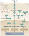
Figure 1. The MAPK and related pathways in thyroid cancer
Shown in the middle of the figure is the classical MAPK pathway leading from an extracellular mitogenic stimulus that activates a receptor tyrosine kinase (RTK) in the cell membrane, to RAS, RAF (shown as BRAF-V600E), MEK and ERK. ERK is activated by phosphorylation (P) and enters the nucleus where it upregulates tumour-promoting genes and downregulates tumour suppressor genes and thyroid iodide-handling genes. On the left side of the figure is the nuclear factor-κB (NF-κB) pathway, in which extracellular stimuli activate the pathway by acting on receptors in the cell membrane, leading to activation of the inhibitor of κB (IκB) kinase (IKK), resulting in the phosphorylation of IκB. Phosphorylated IκB becomes dissociated from NF-κB, which is normally bound with IκB in a complex and sequestered in the cytoplasm. Phosphorylated IκB undergoes ubiquitylation and proteasomal degradation. Free NF-κB then enters the nucleus to promote the expression of tumour-promoting genes. Through an undefined mechanism that is independent of MEK signalling, BRAF-V600E promotes the phosphorylation of IκB and the release of NF-κB, thus activating the NF-κB pathway. Shown on the right side of the figure is the RASSF1–mammalian STE20-like protein kinase 1 (MST1)–forkhead box O3 (FOXO3) pathway. Activated by extracellular pro-apoptotic stimuli through membrane receptors, RASSF1A activates MST1. Activated MST1 then phosphorylates FOXO3 on Ser207. The resulting phosphorylated FOXO3 becomes dissociated from 14-3-3 proteins in the cytoplasm. 14-3-3 proteins undergo proteasomal degradation, and phosphorylated FOXO3 enters the nucleus to promote the expression of pro-apoptotic genes in the FOXO pathway. BRAF-V600E directly interacts with and inhibits MST1 and prevents its activation by RASSF1A, thereby suppressing the pro-apoptotic signalling of the FOXO3 pathway. The downward arrow for the FOXO activities shown in the nucleus indicates this negative effect of BRAF-V600E on pro-apoptotic genes, which are normally upregulated by the RASSF1A–MST1–FOXO3 pathway. The triple independent coupling of BRAF-V600E to the pathways shown here represents a unique and powerful mechanism of thyroid tumorigenesis driven by BRAF-V600E. DAPK1, death-associated protein kinase 1; HIF1A, hypoxia-inducible factor 1α; MMP, matrix metalloproteinase; NIS, sodium–iodide symporter; TGFB1, transforming growth factor β1; TIMP3, tissue inhibitor of metalloproteinases 3; TPO, thyroid peroxidase; TSHR, thyroid-stimulating hormone receptor; TSP1, thrombospondin 1; UPA, urokinase plasminogen activator; UPAR, urokinase plasminogen activator receptor; VEGFA, vascular endothelial growth factor A.
Figure 2
Figure 2. The PI3K–AKT and related pathways in thyroid cancer
Extracellular signals activate receptor tyrosine kinases (RTKs) in the cell membrane, leading to the activation of RAS and subsequent activation of PI3K. Activated PI3K catalyses the conversion of phosphatidylinositol (4,5)-bisphosphate (PIP2) to phosphatidylinositol (3,4,5)-trisphosphate (PIP3). PIP3 activates 3-phosphoinositide-dependent protein kinase 1 (PDK1; also known as PDPK1), which consequently associates with AKT and leads to phosphorylation (P) and activation of AKT by PDK1. Phosphorylated AKT, which is an activated form of AKT, enters the nucleus where it induces tumour-promoting genes. In the cytoplasm, phospho-AKT also activates other signalling molecules or pathways, a prominent one being the mTOR pathway, which has an important role in tumorigenesis by promoting translation. Phospho-AKT can also directly phosphorylate glycogen synthase kinase 3β (GSK3β) and consequently inactivates it. GSK3β normally inhibits β-catenin, thus the effect of phospho-AKT is to relieve GSK3β-mediated suppression of β-catenin. Consequently, β-catenin can enter the nucleus where it promotes the expression of tumour-promoting genes. In the nucleus, phospho-AKT can phosphorylate forkhead box O3 (FOXO3) on its AKT-specific motif. Such phosphorylated FOXO3 is translocated out of the nucleus to the cytoplasm where it binds 14-3-3 proteins to be sequestered in the cytoplasm, thus terminating the pro-apoptotic activities of the FOXO3 pathway. The downward arrow for the FOXO activities in the nucleus in the figure indicates this negative effect of AKT on pro-apoptotic genes in the FOXO pathway, which would otherwise be upregulated by the FOXO3 pathway. This unique coupling of phospho-AKT to the three pathways provides a powerful driving force for thyroid tumorigenesis. The major negative regulatory mechanism of the PI3K–AKT pathway is PTEN, which is a phosphatase that converts PIP3 to PIP2, thus terminating the activation of the pathway. The inset shows the self enhancement mechanism of PI3K–AKT signalling in which genetic-alteration-driven activation of the pathway causes PTEN methylation and silencing with consequent loss of termination of the signalling, thus maintaining the pathway in full and constitutive activation.
Figure 3
Figure 3. Model of the progression of thyroid tumorigenesis driven by the MAPK and PI3K–AKT pathways
Activation of the MAPK pathway by genetic alterations, such as the BRAFV600E mutation, primarily drives the development of papillary thyroid cancer (PTC) from follicular thyroid cells. By contrast, activation of the PI3K–AKT pathway by genetic alterations, such as mutations in RAS, PTEN and PIK3CA, primarily drives the development of follicular thyroid adenoma (FTA) and follicular thyroid cancer (FTC) from follicular thyroid cells. Conversion from FTA to FTC is largely due to increasing activation of the PI3K–AKT pathway. As genetic alterations accumulate and intensify the signalling of each of the two pathways, PTC and FTC can progress to poorly differentiated thyroid cancer (PDTC). When both pathways are fully activated through accumulated genetic alterations, conversion from PDTC to anaplastic thyroid cancer (ATC) is strongly facilitated. It is also possible that PDTC and ATC can both develop de novo directly from follicular thyroid cells, and that ATC can develop from PTC or FTC if appropriate genetic alterations occur. Many secondary molecular alterations also progressively accumulate and synergize with the two pathways in driving the progression of thyroid tumorigenesis, as discussed in the text (not shown in the figure). The increasing number of vertical arrows and colour intensity of the ovals symbolize the increasing genetic alterations and signalling of the two pathways as thyroid tumorigenesis progresses. The figure is modified from REF. © (2007) American Association for Cancer Research.
Figure 4
Figure 4. Iodide-handling machinery in the thyroid cell and its silencing by BRAF-V600E
a | A follicular thyroid cell is shown, which abundantly expresses the molecules involved in the uptake and metabolism of iodide (I), including the sodium–iodide symporter (NIS) in the basal membrane, which transports I coupled with Na+ into the cell from the extracellular compartment. I is then transported into the follicular lumen via pendrin in the apical membrane where it is oxidized by thyroid peroxidase (TPO) and incorporated into tyrosine amino-acid residues in thyroglobulin (TG) to form iodinated TG (TG-I) for the synthesis of thyroid hormone. The whole process is upregulated by cyclic AMP (cAMP) signalling that is triggered by the binding of thyroid-stimulating hormone (TSH) to its receptor (TSHR) in the membrane. With normal expression and function of this system, I is abundantly taken up and accumulated in the follicular thyroid cell and in the follicular lumen. b | BRAF-V600E, through activating the MAPK pathway in thyroid cancer, causes the silencing of thyroid-specific genes and shuts off the iodide-handling machinery. Consequently, I uptake is reduced in the thyroid cell and is sparsely accumulated in the follicular lumen. For simplicity, much molecular detail is omitted.

References

    1. Jemal A, et al. Global cancer statistics. CA Cancer J Clin. 2011;61:69–90. - PubMed
    1. Howlader N, et al. SEER Cancer Statistics Review 1975–2009 (Vintage 2009 Populations) National Cancer Institute. 2012 [online], http://seer.cancer.gov/csr/1975_2009_pops09.
    1. Tuttle RM, et al. Thyroid carcinoma. J Natl Compr Canc Netw. 2010;8:1228–1274. - PubMed
    1. DeLellis RA, Lloyd RV, Heitz PU, Eng C. World Health Organization Classification of Tumours. Pathology And Genetics Of Tumors Of Endocrine Organs. IARC Press; 2004. This book describes the most recent version of World Health Organization classification of thyroid tumours.
    1. Hofstra RM, et al. A mutation in the RET proto-oncogene associated with multiple endocrine neoplasia type 2B and sporadic medullary thyroid carcinoma. Nature. 1994;367:375–376. - PubMed

Publication types