

Morphological characteristics of split-depression fractures of the lateral tibial plateau (Schatzker type II): a computer-tomography-based study
- PMID: 23429973
- PMCID: PMC3631501
- DOI: 10.1007/s00264-013-1825-5
Morphological characteristics of split-depression fractures of the lateral tibial plateau (Schatzker type II): a computer-tomography-based study
Abstract
Purpose: The objective of this study was to evaluate the morphological characteristics of lateral tibial plateau split-depression fractures (Schatzker type II).
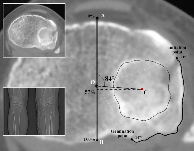
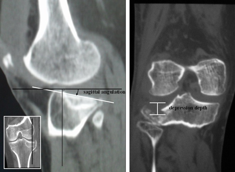
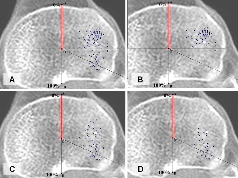
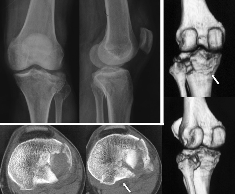
Methods: A retrospective analysis of radiographic and computed tomographic (CT) data of lateral tibial plateau split-depression fractures from January 2009 to December 2010 was conducted in a level 1 trauma centre. The discontinuity arc, angle of depression centre (ADC), anterior-posterior position of articular depression centre (APDC), surface area percentage (SAP), sagittal angulation and depression depth were measured on CT images using the Picture Archiving and Communication System.
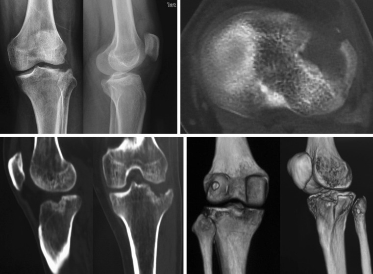
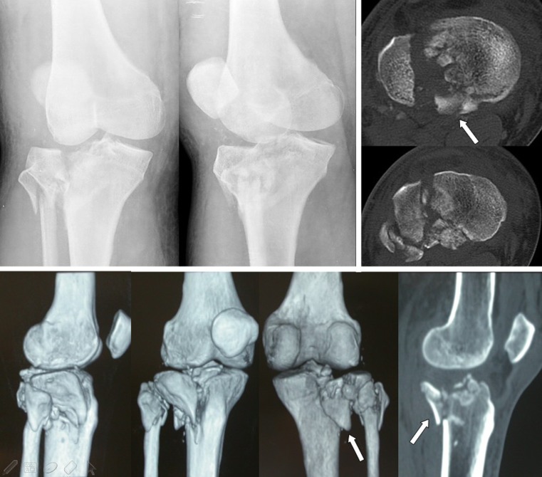
Results: Based on the integrity of posterolateral wall and discontinuity arc, 140 cases of Schatzker type II fracture were divided into two subtypes: intact group (69 cases) and broken group (71 cases). The fracture of the intact group was located in the anterior part of the lateral plateau, the average ADC was 72.13°, APDC was 43.25 % of sagittal diameter, SAP was 22.16 % of total plateau, sagittal angulation was 6.59°and depression depth was 10.80 mm. Of the broken group, the average ADC, APDC, SAP, sagittal angulation and depression depth was 92.45°, 61.84 %, 22.63 %, 9.00° and 10.78 mm, respectively. Forty-seven cases in the broken group had a posterolateral fragment and 15 cases among them had articular depression centered in the posterolateral region. The difference in ADC, APDC and sagittal angulation between the two groups was statistically significant (p < 0.05), while no significant difference was observed for SAP and depression depth.
Conclusions: Of all the 140 cases in this study, 10.7 % are located in the posterolateral region. An appropriate operative approach and fixation method should be selected based on the individual morphological characteristics of lateral plateau fractures.
Figures
Comment in
-
Posterolateral fragment characteristics in tibial plateau fractures.Int Orthop. 2014 Mar;38(3):681-2. doi: 10.1007/s00264-013-2248-z. Epub 2014 Jan 3. Int Orthop. 2014. PMID: 24384937 Free PMC article. No abstract available.
References
-
- Kennedy JC, Bailey WH. Experimental tibial-plateau fractures. Studies of the mechanism and a classification. J Bone Joint Surg Am. 1968;50(8):1522–1534. - PubMed
-
- Marsh JL, Slongo TF, Agel J, Broderick JS, Creevey W, DeCoster TA, Prokuski L, Sirkin MS, Ziran B, Henley B, Audigé L. Fracture and Dislocation Classification Compendium - 2007: Orthopaedic Trauma Association Classification, Database and Outcomes Committee. J Orthop Trauma. 2007;21(10 Su ppl):S1–S133. doi: 10.1097/00005131-200711101-00001. - DOI - PubMed
-
- Schatzker J, McBroom R, Bruce D. The tibial plateau fracture. The Toronto experience 1968-1975. Clin Orthop Relat Res. 1979;138:94–104. - PubMed
-
- Zhu Y, Yang G, Luo CF, Smith WR, Hu CF, Gao H, Zhong B, Zeng BF. Computed tomography-based Three-Column Classification in tibial plateau fractures: introduction of its utility and assessment of its reproducibility. J Trauma Acute Care Surg. 2012;73(3):731–737. doi: 10.1097/TA.0b013e31825c17e7. - DOI - PubMed
MeSH terms
LinkOut - more resources
Full Text Sources
Other Literature Sources
Medical
Miscellaneous
