. 2013 Mar 29;288(13):9249-60.
doi: 10.1074/jbc.M112.417725. Epub 2013 Feb 19.

Variable electron transfer pathways in an amphibian cryptochrome: tryptophan versus tyrosine-based radical pairs

Affiliations

Variable electron transfer pathways in an amphibian cryptochrome: tryptophan versus tyrosine-based radical pairs

Till Biskup et al. J Biol Chem. .

Abstract

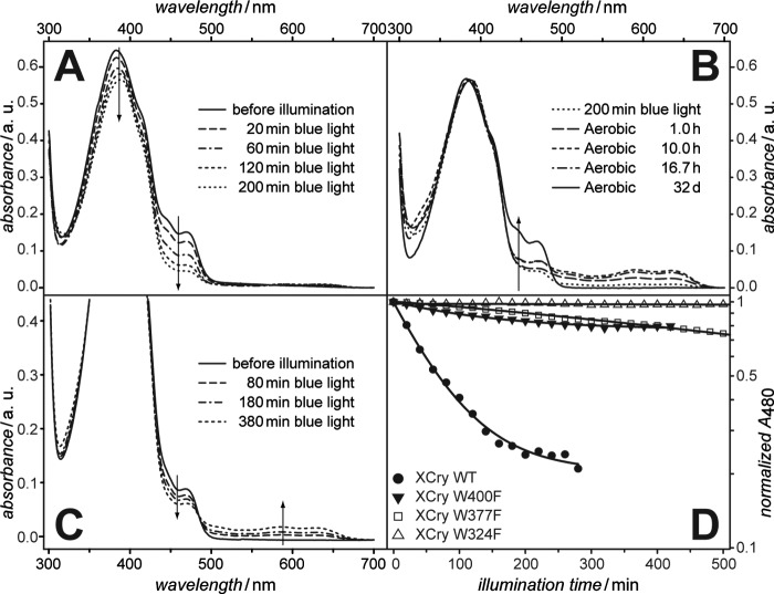
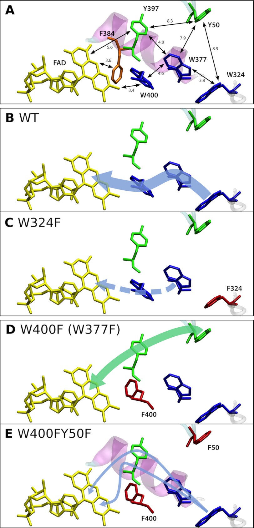
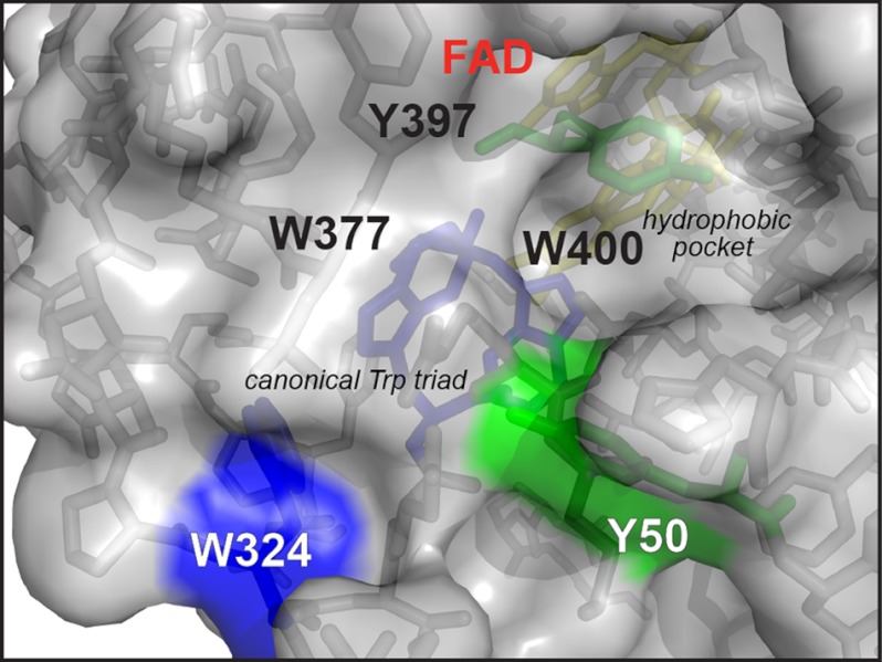
Electron transfer reactions play vital roles in many biological processes. Very often the transfer of charge(s) proceeds stepwise over large distances involving several amino acid residues. By using time-resolved electron paramagnetic resonance and optical spectroscopy, we have studied the mechanism of light-induced reduction of the FAD cofactor of cryptochrome/photolyase family proteins. In this study, we demonstrate that electron abstraction from a nearby amino acid by the excited FAD triggers further electron transfer steps even if the conserved chain of three tryptophans, known to be an effective electron transfer pathway in these proteins, is blocked. Furthermore, we were able to characterize this secondary electron transfer pathway and identify the amino acid partner of the resulting flavin-amino acid radical pair as a tyrosine located at the protein surface. This alternative electron transfer pathway could explain why interrupting the conserved tryptophan triad does not necessarily alter photoreactions of cryptochromes in vivo. Taken together, our results demonstrate that light-induced electron transfer is a robust property of cryptochromes and more intricate than commonly anticipated.


Figures

FIGURE 1.
FIGURE 1.
Blue-light induced spectral changes in the optical absorptions of WT and mutant XCrys. All measurements were performed in the presence of 10 mm EDTA (280 K). A, spectral changes in the optical absorptions of WT XCry after the indicated illumination times under anaerobic conditions. B, spectral changes in the reoxidation process by atmospheric oxygen in the dark. C, representative spectral changes in the optical absorption of XCry W400F after the indicated illumination times under anaerobic conditions. D, comparison of the photoreduction speed at 480 nm of WT XCry, W324F, W377F, and W400F performed under anaerobic conditions. Fits (black lines) were calculated using the reaction scheme described under “Experimental Procedures.”
FIGURE 2.
FIGURE 2.
Transient optical spectroscopy of WT XCry. A, difference absorption spectra of WT XCry in the wavelength region between 370 and 700 nm recorded at the indicated times after pulsed laser excitation. Each spectrum is the average of 10 time points (corresponding to an integration window of 50 ns for spectra taken at 150 ns and 2 μs and a window of 5 μs for spectra taken at 20, 100, and 200 μs). B, selected transients (410, 450, 510, and 582 nm) of WT XCry. Each time profile is the average of two acquisitions recorded with a shot repetition rate of 0.016 Hz. All spectra were recorded at 279 K. C, species-associated difference spectrum components of WT XCry corresponding to the kinetic model shown in D. For further details, see text.
FIGURE 3.
FIGURE 3.
TREPR signals generated by pulsed laser excitation (460 nm; 1.25/1 Hz pulse repetition rate; 4 mJ pulse energy) of XCry (A) WT, (B) W324F, (C) W400F and W377F (upper and lower green trace, respectively), (D) Y50F/W400F, and (E) W324F/W400F, recorded at 274/270 K centered at 500 ns after the laser pulse with an integration window of 200 ns in direct detection mode (integrated amplitudes with A, enhanced absorption, and E, emission). Please note that spectra A and B were taken from Ref. and mutants Y50F/W400F and W324F/W400F were measured with a different setup and thus, their signal strength cannot be directly compared with that of the other XCry samples. Instrument settings: 9.69 GHz; microwave power: 2 mW. Each data point represents the average of 60 acquisitions (100 for Y50F/W400F and W324F/W400F, respectively) recorded with a detection bandwidth of 100 MHz (25 MHz for Y50F/W400F and W324F/W400F, respectively). The dashed gray curves show spectral simulations for the FAD···Tyr-50 RP made with the following parameters: gFAD = (2.00431, 2.00360, and 2.00217); gTyr-50 = (2.00767, 2.00438, and 2.00219); dipolar interaction, D = −0.51 mT; exchange interaction, J = +0.24 mT. For details, see supplemental data and supplemental Table S2.
FIGURE 4.
FIGURE 4.
Proposed electron transfer pathways in WT and mutant XCry. A, selected residues of the XCry model structure showing the FAD cofactor (yellow) and the conserved Trp triad (blue). The proposed alternative ET pathway along Tyr-397 and Tyr-50 is shown in green. B to E, schematic representation showing the proposed ET pathways for each of the investigated XCry mutants (dashed pathway in panel C is proposed but could not be observed experimentally). Please note that the ET pathway in panel E remains highly ambiguous. For further details, see text.
FIGURE 5.
FIGURE 5.
Surface accessibility of the aromatic amino acids potentially acting as terminal electron donors. Trp-324 is depicted in blue and Tyr-50 is depicted in green.

References

    1. Ahmad M., Cashmore A. R. (1993) HY4 gene of A. thaliana encodes a protein with characteristics of a blue-light photoreceptor. Nature 366, 162–166 - PubMed
    1. Chaves I., Pokorny R., Byrdin M., Hoang N., Ritz T., Brettel K., Essen L. O., van der Horst G. T., Batschauer A., Ahmad M. (2011) The cryptochromes. Blue light photoreceptors in plants and animals. Annu. Rev. Plant Biol. 62, 335–364 - PubMed
    1. Losi A., Gärtner W. (2011) Old chromophores, new photoactivation paradigms, trendy applications. Flavins in blue light-sensing photoreceptors. Photochem. Photobiol. 87, 491–510 - PubMed
    1. Zoltowski B. D., Gardner K. H. (2011) Tripping the light fantastic. Blue-light photoreceptors as examples of environmentally modulated protein–protein interactions. Biochemistry 50, 4–16 - PMC - PubMed
    1. Essen L. O. (2006) Photolyases and cryptochromes. Common mechanisms of DNA repair and light-driven signaling? Curr. Opin. Struct. Biol. 16, 51–59 - PubMed

Publication types

LinkOut - more resources