. 2013 Apr 5;288(14):9755-9766.
doi: 10.1074/jbc.M112.446435. Epub 2013 Feb 21.

Three-dimensional structure of Saccharomyces invertase: role of a non-catalytic domain in oligomerization and substrate specificity

Affiliations

Three-dimensional structure of Saccharomyces invertase: role of a non-catalytic domain in oligomerization and substrate specificity

M Angela Sainz-Polo et al. J Biol Chem. .

Abstract

Invertase is an enzyme that is widely distributed among plants and microorganisms and that catalyzes the hydrolysis of the disaccharide sucrose into glucose and fructose. Despite the important physiological role of Saccharomyces invertase (SInv) and the historical relevance of this enzyme as a model in early biochemical studies, its structure had not yet been solved. We report here the crystal structure of recombinant SInv at 3.3 Å resolution showing that the enzyme folds into the catalytic β-propeller and β-sandwich domains characteristic of GH32 enzymes. However, SInv displays an unusual quaternary structure. Monomers associate in two different kinds of dimers, which are in turn assembled into an octamer, best described as a tetramer of dimers. Dimerization plays a determinant role in substrate specificity because this assembly sets steric constraints that limit the access to the active site of oligosaccharides of more than four units. Comparative analysis of GH32 enzymes showed that formation of the SInv octamer occurs through a β-sheet extension that seems unique to this enzyme. Interaction between dimers is determined by a short amino acid sequence at the beginning of the β-sandwich domain. Our results highlight the role of the non-catalytic domain in fine-tuning substrate specificity and thus supplement our knowledge of the activity of this important family of enzymes. In turn, this gives a deeper insight into the structural features that rule modularity and protein-carbohydrate recognition.


Figures
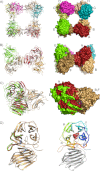
FIGURE 1.
FIGURE 1.
Structure of octameric SInv. a, view of the SInv octamer in ribbon (left) and solvent-accessible surface (right) representations, showing each subunit in a different color. In b, the octamer is rotated 90°, illustrating that it can be best described as a tetramer of two different kinds of dimers, AB/CD and EF/HG, which are compared by superimposing subunit F on subunit B in c. Then, a rotation of 15° would be necessary to bring out subunit E into the A position. Three regions of the polypeptide chain act as hinges (DynDom server), which are colored blue in d (left). The different dimer associations produce local conformational differences at the dimer interface in specific regions represented in red (A, B, C, and D) and green (E, F, G, and H). SInv folds into two domains, a catalytic β-propeller that is colored according to its five blades (I–V) and a C-terminal β-sandwich domain formed by two antiparallel six-stranded β-sheets (right). The putative position of a 1-kestose substrate molecule shown in c and d is inferred from structural superposition of SInv on the Cichorium intybus fructan 1-exohydrolase·1-kestose complex (Protein Data Bank code 2AEZ) to point out the active site cavity.
FIGURE 2.
FIGURE 2.
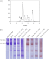
Analysis of SInv oligomeric state. a, size exclusion analysis of SInv. A sample of purified enzyme (2 mg/ml) in 0.05 m phosphate buffer (pH 7) and 150 mm NaCl was injected onto a Superdex 200 10/300 GL column coupled to an ÅKTA purifier system (GE Healthcare) previously equilibrated with the same buffer. Elution was carried out at a flow rate of 0.5 ml/min for 1.5 column volumes (solid line). SInv eluted mainly as a peak of ∼430 kDa (peak 1), indicative of an octameric structure. A shoulder of this peak at lower elution volumes, highlighted with an arrow (peak 2), probably corresponds to higher molecular mass aggregates. Calibration of the column was performed with molecular mass standards (Bio-Rad catalog no. 151-1901) eluted under the same conditions (dotted line): peak a, thyroglobulin (670 kDa); peak b, γ-globulin (158 kDa); peak c, ovalbumin (44 kDa); peak d, myoglobin (17 kDa); and peak e, vitamin B12 (1.35 kDa). b, nondenaturing PAGE analysis of purified SInv performed on 6% polyacrylamide gels. Gels were stained with Coomassie Blue (left), or alternatively, invertase activity was detected by incubating the gels in sucrose and subsequently staining with 1% (w/v) 2,3,5-triphenyltetrazolium chloride in 0.25 m NaOH (right) as described previously (22). Oligomers lower than the octamer appeared after heating SInv at 47 °C or incubating with urea. These treatments also decreased the proportion of the sample that formed higher aggregates. Other external conditions, such as addition of NaCl (0.1, 0.5, and 1 m), dilution (1:3, 1:5, and 1:10), or varying pH (4.5, 7.5, and 8.5), did not alter the pattern of untreated SInv (not shown).
FIGURE 3.
FIGURE 3.
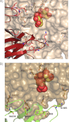
Dimer interface at the active site. The octameric SInv active site interfaces are detailed, keeping the same color pattern in Fig. 1c, with one subunit being shown in ribbon representation for clarity. a, the AB/CD dimers are tightly made by interactions among both their catalytic and β-sandwich domains. The base of the catalytic pocket is additionally lined by hydrophobic interactions through Phe-388 and Phe-296. b, by contrast, the EF/GH dimers interact only through their β-sandwich domains. In addition, the catalytic pocket is also paved by a new salt bridge formed between Asp-45 and Lys-385 from the β-sandwich domain, which lines the cavity. A putative 1-kestose molecule is shown in spherical representation (inferred as explained in the legend to Fig. 1).
FIGURE 4.
FIGURE 4.
Intermolecular β-sheet. Both sets of dimers assemble through a similar interface that involves the extension of their corresponding β-sheets between each β-sandwich domain. a, view of the internal part at the octamer interface showing that the hydrogen bond pattern centered on β2 corresponds to a regular β-sheet. b, in contrast, the external wall is defined by polar links between the side chains of the residues at β1.
FIGURE 5.
FIGURE 5.
Structural differences between SInv and SoFfase. a, structural alignment of the SoFfase subunit (slate) and the SInv monomer (red) shows well conserved catalytic domains but larger differences in the β-sandwich domain, mostly at strands β1 and β2, at the base of the β-sandwich domain, which can be attributed to the interactions of the β-sandwich elements in the SInv octamer. b, the β-sandwich of an SInv subunit is shown in surface representation to highlight the effect of the different arrangement in the active site (magnified) at the dimer interface. The putative 1-kestose position is shown in spherical representation (inferred as explained in the legend to Fig. 1).
FIGURE 6.
FIGURE 6.
Specificity of SInv. Shown are stereo views of the SInv active site of the AB/CD (a) and EF/GH (b) dimers compared with the SoFfase dimer (c). The putative position of the substrate 1-kestose (inferred as explained in the legend to Fig. 1) is shown in a and b. A fructosylnystose molecule (beige) found in the reported E230A SoFfase complex (Protein Data Bank code 3U75) shown in c could enter into the EF/GH active site pocket (b), whereas the narrow entrance at the AB/CD interface (a) would prevent binding of extended fructans. Moreover, in SInv, the short chain residues surrounding the cavity outline a more rigid and therefore less flexible active site to accommodate long and polymeric oligosaccharides. d, structural alignment of SoFfase (slate) in the SInv catalytic pockets (ABCD (red) and EFGH (green)) showing the nucleophile (Asp-22), the intermediate stabilizer (Asp-151), and the acid/base catalyst (Glu-203). The putative positions of the transfructosylation products 1-kestose and 6-kestose are shown in cyan and purple, respectively. The position of 6-kestose has been inferred from superposition of its coordinates extracted from the Cambridge Structural Database (CSD; refcode CELGIC) onto the fructose of docked 1-kestose at subsite −1. The recognition scheme of the donor sucrose moiety giving each product within the AB/CD dimers would be the same as that described for SoFfase, as explained under “Results.”
FIGURE 7.
FIGURE 7.
Model of extracellular SInv. Shown is a putative model of secreted octameric SInv built manually from the AB dimers, with retention of the intermolecular β-sheet. This model reproduces the electron micrographs reported for this isoform (10, 11), which showed rectangles slightly open in one side.
FIGURE 8.
FIGURE 8.
Sequence conservation within GH32 family members from yeast. The SInv sequence is aligned with other yeast enzymes: Kluyveromyces marxianus inulinase (KmInu), SoFfase, Candida sp KRF1 inulinase (CaInu), and Schizosaccharomyces pombe invertase (SpInv). Conserved regions are highlighted (44). The regions of the catalytic domain involved in making the dimer interface are shown in a, whereas the β-sandwich domain is shown in b. Stars specify residues involved in polar links within the AB/CD dimer interface. Inverted triangles indicate residues that make polar links between the β-sandwich domains from contiguous subunits responsible for the formation of the intermolecular β-sheets. Solid circles are potential N-glycosylation sites in extracellular SInv.

References

    1. Barnett J. A. (2003) Beginnings of microbiology and biochemistry: the contribution of yeast research. Microbiology 149, 557–567 - PubMed
    1. Michaelis L., Menten M. L. (1913) The Kinetics of Invertase Action. Biochem. Z. 49, 333–369
    1. de la Fuente G., Sols A. (1962) Transport of sugars in yeasts. II. Mechanisms of utilization of disaccharides and related glycosides. Biochim. Biophys. Acta 56, 49–62 - PubMed
    1. Gascón S., Lampen J. O. (1968) Purification of the internal invertase of yeast. J. Biol. Chem. 243, 1567–1572 - PubMed
    1. Gascón S., Neumann N. P., Lampen J. O. (1968) Comparative study of the properties of the purified internal and external invertases from yeast. J. Biol. Chem. 243, 1573–1577 - PubMed

Publication types

MeSH terms

Associated data