Review
. 2013 Feb 15;27(4):355-71.
doi: 10.1101/gad.210773.112.

The Hippo pathway: regulators and regulations

Affiliations
Review

The Hippo pathway: regulators and regulations

Fa-Xing Yu et al. Genes Dev. .

Abstract

Control of cell number is crucial in animal development and tissue homeostasis, and its dysregulation may result in tumor formation or organ degeneration. The Hippo pathway in both Drosophila and mammals regulates cell number by modulating cell proliferation, cell death, and cell differentiation. Recently, numerous upstream components involved in the Hippo pathway have been identified, such as cell polarity, mechanotransduction, and G-protein-coupled receptor (GPCR) signaling. Actin cytoskeleton or cellular tension appears to be the master mediator that integrates and transmits upstream signals to the core Hippo signaling cascade. Here, we review regulatory mechanisms of the Hippo pathway and discuss potential implications involved in different physiological and pathological conditions.


Figures
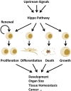
Figure 1.
Figure 1.
Implications of the Hippo pathway in cell biology. The Hippo pathway modulates cell proliferation, differentiation, growth, and death. The coordination of different cellular processes by the Hippo pathway may contribute to diverse physiological and pathological conditions such as development, tissue homeostasis, and tumorigenesis.
Figure 2.
Figure 2.
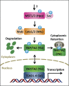
The core Hippo pathway. MST1/2 phosphorylates Sav, Lats1/2, and Mob; Lats1/2 phosphorylates YAP/TAZ; and phosphorylated YAP/TAZ interacts with 14-3-3 and results in cytoplasmic retention. Moreover, YAP/TAZ phosphorylation leads to protein degradation. When dephosphorylated, YAP/TAZ enter nuclei and induce gene transcription by interacting with transcription factors TEAD1–4. Drosophila orthologs for these core components are shown in brackets.
Figure 3.
Figure 3.
Regulatory mechanisms for the Hippo pathway. Regulation of the Hippo pathway by apical–basal polarity (A), PCP (B), mechanical cues and GPCR signaling (C), and actin cytoskeleton (D). Arrowed or blunted ends indicate activation or inhibition, respectively. Dashed lines indicate indirect or unknown mechanisms. Red lines in D represent actin filaments.
Figure 4.
Figure 4.
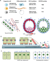
YAP/TAZ activity under different physiological and pathological conditions. (A) YAP/TAZ activity in cell differentiation. YAP/TAZ activity is regulated by cell density, ECM, and GPCR signaling. (B) Cells close to a wound may experience different mechanical forces and a higher concentration of GPCR ligands from blood vessels. This may induce YAP/TAZ activity and promote wound healing or tissue regeneration. (C) In a blastocyst, cells in the trophectoderm (TE) and inner cell mass (ICM) have distinct YAP localization and F-actin/tension distribution. (D) Disruption of cellular junctions may induce mechanical stress and result in changes in cell morphology, which can enrich YAP/TAZ in nuclei under these conditions. Activated YAP/TAZ may promote epithelial–mesenchymal transition (EMT) and cell migration. Cells that have escaped from epithelium may also encounter more GPCR ligands. Red lines represent actin filaments. (E) Aberrant GPCR signaling may activate YAP/TAZ. Elevated GPCR expression or activating Gα mutations may induce YAP/TAZ nuclei localization and activation and result in hyperproliferation that may contribute to cancer development. YAP/TAZ localization is represented by green in BE.

References

    1. Alarcon C, Zaromytidou AI, Xi Q, Gao S, Yu J, Fujisawa S, Barlas A, Miller AN, Manova-Todorova K, Macias MJ, et al. 2009. Nuclear CDKs drive Smad transcriptional activation and turnover in BMP and TGF-β pathways. Cell 139: 757–769 - PMC - PubMed
    1. Azzolin L, Zanconato F, Bresolin S, Forcato M, Basso G, Bicciato S, Cordenonsi M, Piccolo S 2012. Role of TAZ as mediator of Wnt signaling. Cell 151: 1443–1456 - PubMed
    1. Balazs L, Okolicany J, Ferrebee M, Tolley B, Tigyi G 2001. Topical application of the phospholipid growth factor lysophosphatidic acid promotes wound healing in vivo. Am J Physiol Regul Integr Comp Physiol 280: R466–R472 - PubMed
    1. Bao Y, Nakagawa K, Yang Z, Ikeda M, Withanage K, Ishigami-Yuasa M, Okuno Y, Hata S, Nishina H, Hata Y 2011. A cell-based assay to screen stimulators of the Hippo pathway reveals the inhibitory effect of dobutamine on the YAP-dependent gene transcription. J Biochem 150: 199–208 - PubMed
    1. Baum B, Georgiou M 2011. Dynamics of adherens junctions in epithelial establishment, maintenance, and remodeling. J Cell Biol 192: 907–917 - PMC - PubMed

Publication types

MeSH terms

Substances