. 2013 Mar 5;110(10):4087-92.
doi: 10.1073/pnas.1218424110. Epub 2013 Feb 19.

Large α-synuclein oligomers inhibit neuronal SNARE-mediated vesicle docking

Affiliations

Large α-synuclein oligomers inhibit neuronal SNARE-mediated vesicle docking

Bong-Kyu Choi et al. Proc Natl Acad Sci U S A. .

Abstract

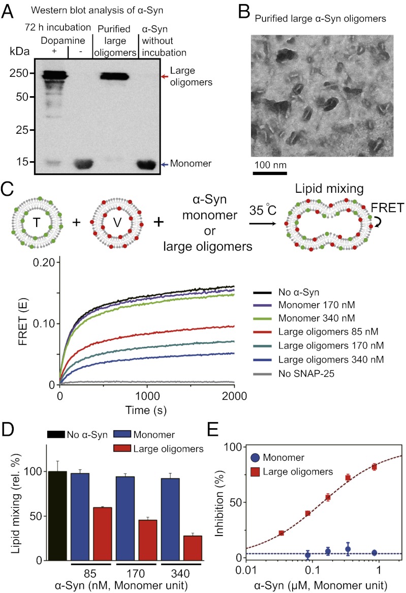
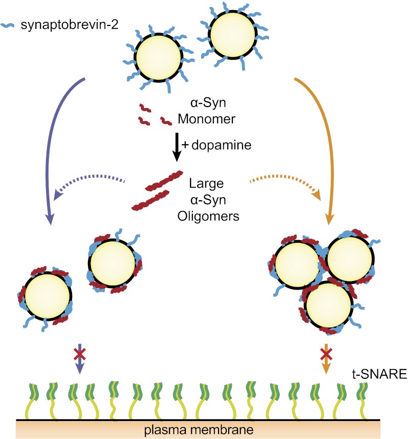
Parkinson disease and dementia with Lewy bodies are featured with the formation of Lewy bodies composed mostly of α-synuclein (α-Syn) in the brain. Although evidence indicates that the large oligomeric or protofibril forms of α-Syn are neurotoxic agents, the detailed mechanisms of the toxic functions of the oligomers remain unclear. Here, we show that large α-Syn oligomers efficiently inhibit neuronal SNARE-mediated vesicle lipid mixing. Large α-Syn oligomers preferentially bind to the N-terminal domain of a vesicular SNARE protein, synaptobrevin-2, which blocks SNARE-mediated lipid mixing by preventing SNARE complex formation. In sharp contrast, the α-Syn monomer has a negligible effect on lipid mixing even with a 30-fold excess compared with the case of large α-Syn oligomers. Thus, the results suggest that large α-Syn oligomers function as inhibitors of dopamine release, which thus provides a clue, at the molecular level, to their neurotoxicity.


Conflict of interest statement

The authors declare no conflict of interest.

Figures

Fig. 1.
Fig. 1.
Dopamine-induced large α-Syn oligomers and their inhibitory effect on SNARE-mediated lipid mixing. (A) Western blot of the α-Syn monomers and oligomers. First lane, α-Syn oligomers generated by incubating 10 μM α-Syn with 100 μM dopamine at 37 °C for 72 h. Second lane, a control without dopamine. Third lane, purified large α-Syn oligomers (Fig. S1). Fourth lane, α-Syn monomers. (B) Typical electron micrograph of purified large α-Syn oligomers. Rod-shaped large α-Syn oligomers are dominant. The average length and width are estimated to be 37 nm and 5 nm, respectively (Fig. S2). (C) The effect of large α-Syn oligomers on an in vitro lipid-mixing assay. When t-vesicles doped with donor dyes [reconstituted with t-SNAREs [T]) and v-vesicles doped with acceptor dyes [reconstituted synaptobrevin-2 (V)] are hemifused or fused together, lipid mixing between two vesicles occurs and increases the FRET signal. No α-Syn: T and V (20 μM in lipid concentration) were mixed together at 35 °C without α-Syn. No SNAP-25: a lipid mixing control without SNAP-25 in T. When the α-Syn monomers were added to the TV mixture, no inhibition of lipid mixing was observed (170 nM, violet line; 340 nM, light green line). When large α-Syn oligomers were added to the TV mixture, a significant reduction was observed in lipid mixing (85 nM, red line; 170 nM, dark green line; 340 nM, blue line; all concentrations in monomer units). (D) Relative percentages of lipid mixing at 1,800 s from C. (E) Percentages of lipid-mixing inhibition at various α-Syn concentrations. Blue ●, monomers; red ▪, large oligomers. Error bars were obtained from three independent experiments.
Fig. 2.
Fig. 2.
Large α-Syn oligomers inhibit lipid mixing by binding to the N-terminal domain of synaptobrevin-2. (A) Test of large α-Syn oligomer binding to phospholipids. Protein-free vesicles (F) (50 nm in diameter) were added to the 20 μM TV mixture in the presence of 170 nM large α-Syn oligomers (blue line, no F; red line, 20 μM F; and green line, 40 μM F, in lipid concentrations). Addition of F has no effect on lipid-mixing inhibition. Results are summarized in the bar graph. (B) Large α-Syn oligomers binding to SNARE-carrying proteoliposomes. While keeping the concentration of large α-Syn oligomers the same, the amount of TV mixture was varied from 20 μM to 60 μM (in lipid concentration) (± SD, n = 3). (C) Coflotation assay for large α-Syn oligomer binding to vesicles. (Left) Schematic description of the coflotation assay. T, V, ntV, or F was incubated with large α-Syn oligomers, respectively, and then vesicle-bound large α-Syn oligomers were separated from unbound large α-Syn oligomers. Here, ntV denotes v-vesicles reconstituted with N-terminal truncated synaptobrevin-2 (nt-synaptobrevin-2, amino acids 29–116). (Right) The amounts of vesicle-bound large α-Syn oligomers were quantified by Western blot (± SD, n = 3). (D) The effect of large α-Syn oligomers on lipid mixing reconstituted with nt-synaptobrevin-2. Bar graphs were obtained from three independent measurements.
Fig. 3.
Fig. 3.
Single-vesicle assay for inhibition of lipid mixing by large α-Syn oligomers. (A) Schematic description of single-vesicle lipid-mixing assay by ALEX. Sufficient dilution to 100 pM vesicle concentration ensures that only one vesicle passes through the excitation volume at a given time. The detected fluorescent emissions result in fluorescence time traces (SI Text). (B) Typical fluorescence time traces of vesicles obtained by ALEX. ALEX generates three types of fluorescence emissions for a vesicle: formula image
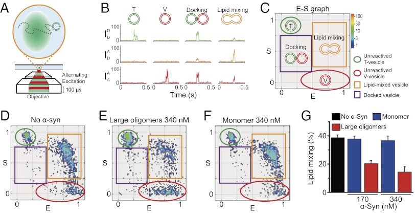
, emission of donor dye [1,1′-dioctadecyl-3,3,3′,3′-tetramethylindocarbocyanine perchlorate (DiI)] excited by donor-excitation laser (green line); formula image, emission of acceptor dye [1,1′-dioctadecyl-3,3,3′,3′-tetramethylindodicarbocyanine perchlorate (DiD)] excited by donor-excitation laser, which is FRET signal (orange line); and formula image, emission of acceptor dye excited by acceptor-excitation laser (red line). Depending on the reaction status, unreacted T and V, docked, and lipid-mixed vesicles have different sets of three fluorescence intensities. (C) Schematic description of the 2D E (FRET efficiency)–S (sorting number) graph. Three fluorescent intensities of a vesicle from the time traces in B are used to calculate E and S (SI Text). Unreacted, docked, and lipid-mixed vesicles locate at different areas in the ES graph: T (green oblique), V (red oblique), docked vesicle (purple box), and lipid-mixed vesicle (orange box). (D–F) 2D ES graphs obtained by ALEX. Each dot denotes a vesicle. (D) Without α-Syn. (E) In the presence of 340 nM of large α-Syn oligomers. (F) In the presence of 340 nM α-Syn monomer. (G) Fractions of lipid-mixed vesicles measured from ALEX (± SD, n = 3).
Fig. 4.
Fig. 4.
Large α-Syn oligomers transduced into PC12 cells reduced exocytosis. (A) The delivery of α-Syn monomers and large oligomers into PC12 cells was confirmed by Western blot. Cell lysates, 50 μg, were applied to the Western blot analysis and probed with anti–α-Syn antibody (Upper). Actin levels were measured to confirm the equal amount of protein loading (Lower). (B) The effect of large α-Syn oligomers on exocytosis in PC12 cells. After transfecting α-Syn into the cells, the amount of [14C]-acetylcholine released by the depolarization of docked vesicles with a solution of high-K+ concentration was measured. Gray bar, without α-Syn; blue and red bars, transduced with α-Syn monomer and large oligomers, respectively (***P < 0.005). Cell viability after transfecting α-Syn was measured by the MTT method (green ▪). (C) Controls without transfection reagents. PC12 cells were incubated with the α-Syn monomers or large oligomers without the treatment of transfecting reagents. All error bars were obtained from five independent measurements.
Fig. 5.
Fig. 5.
A model for large α-Syn oligomers’ inhibition effect on exocytosis. Oxidative stress induces oxidants of dopamine that accelerate the formation of large α-Syn oligomers. Large α-Syn oligomers bind to the N-terminal domain of synaptobrevin-2, which blocks SNARE complex formation. Large α-Syn oligomers may sequester most of the synaptobrevin-2 using multiple binding sites for synaptobrevin-2 on vesicles; thus, less synaptobrevin-2 is available for SNARE complex formation. Alternatively, multiple synaptobrevin-2 binding sites on a large α-Syn oligomer bind synaptobrevin-2 proteins from several vesicles to induce vesicle clustering, which limits the number of v-vesicles available for docking.

References

    1. Iwai A, et al. The precursor protein of non-A beta component of Alzheimer’s disease amyloid is a presynaptic protein of the central nervous system. Neuron. 1995;14(2):467–475. - PubMed
    1. Clayton DF, George JM. The synucleins: A family of proteins involved in synaptic function, plasticity, neurodegeneration and disease. Trends Neurosci. 1998;21(6):249–254. - PubMed
    1. Spillantini MG, et al. Alpha-synuclein in Lewy bodies. Nature. 1997;388(6645):839–840. - PubMed
    1. Irizarry MC, et al. Nigral and cortical Lewy bodies and dystrophic nigral neurites in Parkinson’s disease and cortical Lewy body disease contain alpha-synuclein immunoreactivity. J Neuropathol Exp Neurol. 1998;57(4):334–337. - PubMed
    1. Polymeropoulos MH, et al. Mutation in the alpha-synuclein gene identified in families with Parkinson’s disease. Science. 1997;276(5321):2045–2047. - PubMed

Publication types

MeSH terms

LinkOut - more resources