. 2013 Mar 5;110(10):4045-50.
doi: 10.1073/pnas.1211284110. Epub 2013 Feb 19.

Functional genomic screening identifies dual leucine zipper kinase as a key mediator of retinal ganglion cell death

Affiliations

Functional genomic screening identifies dual leucine zipper kinase as a key mediator of retinal ganglion cell death

Derek S Welsbie et al. Proc Natl Acad Sci U S A. .

Abstract

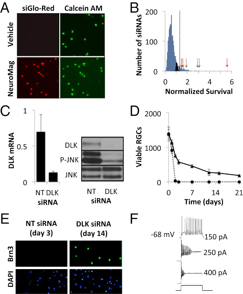
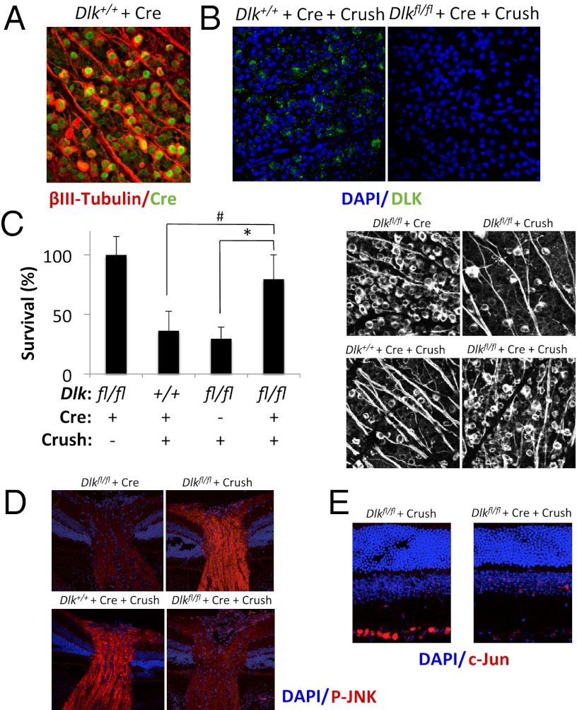
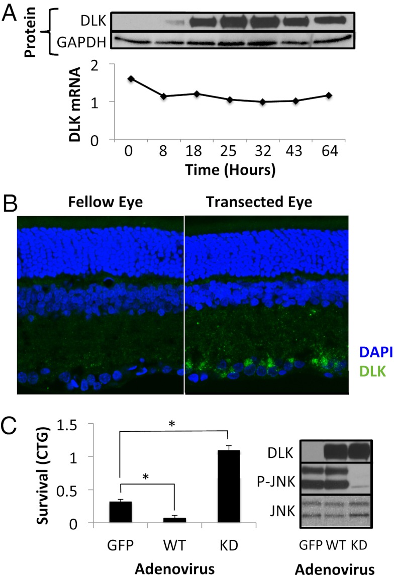
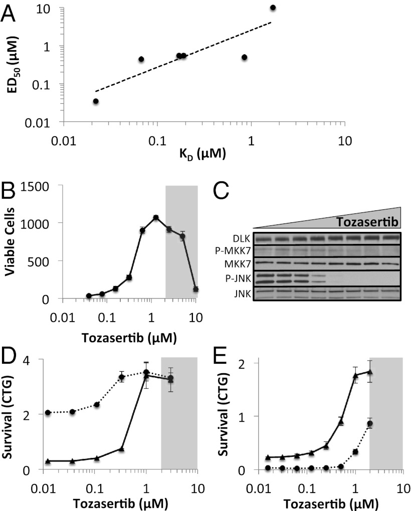
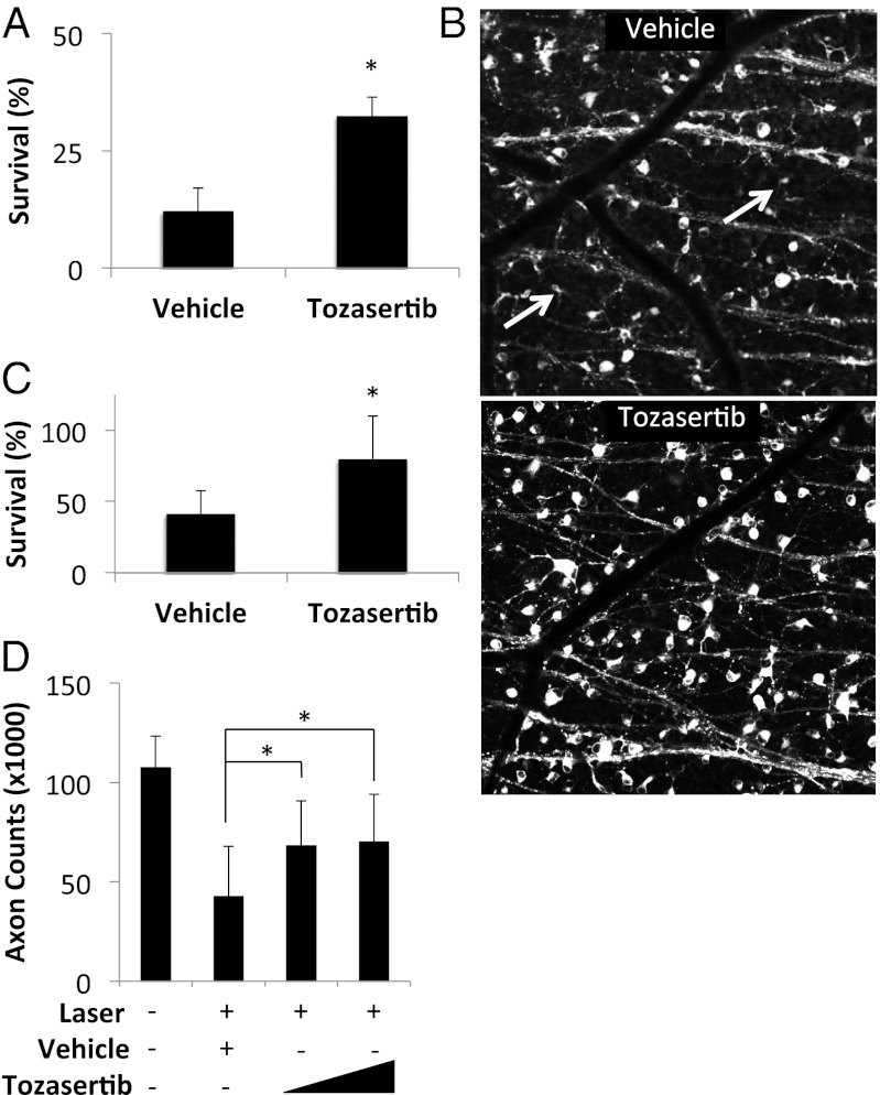
Glaucoma, a major cause of blindness worldwide, is a neurodegenerative optic neuropathy in which vision loss is caused by loss of retinal ganglion cells (RGCs). To better define the pathways mediating RGC death and identify targets for the development of neuroprotective drugs, we developed a high-throughput RNA interference screen with primary RGCs and used it to screen the full mouse kinome. The screen identified dual leucine zipper kinase (DLK) as a key neuroprotective target in RGCs. In cultured RGCs, DLK signaling is both necessary and sufficient for cell death. DLK undergoes robust posttranscriptional up-regulation in response to axonal injury in vitro and in vivo. Using a conditional knockout approach, we confirmed that DLK is required for RGC JNK activation and cell death in a rodent model of optic neuropathy. In addition, tozasertib, a small molecule protein kinase inhibitor with activity against DLK, protects RGCs from cell death in rodent glaucoma and traumatic optic neuropathy models. Together, our results establish a previously undescribed drug/drug target combination in glaucoma, identify an early marker of RGC injury, and provide a starting point for the development of more specific neuroprotective DLK inhibitors for the treatment of glaucoma, nonglaucomatous forms of optic neuropathy, and perhaps other CNS neurodegenerations.


Conflict of interest statement

Conflict of interest statement: D.S.W., Z.Y., S.E.M., H.Q., C.A.B., and D.J.Z. are inventors on patents related to the work described; these patents are being managed by The Johns Hopkins University School of Medicine. W.W.H. and the University of Florida have a financial interest in the use of adeno-associated virus therapies and own equity in a company (AGTC Inc.) that might, in the future, commercialize some aspects of this work.

Figures

Fig. 1.
Fig. 1.
Identification of DLK as a mediator of cell death in RGCs. (A) RGCs were transfected at the time of immunopanning with a fluorescently labeled siRNA (siGLO-Red, Dharmacon) in the presence or absence of the magnetic nanoparticle, NeuroMag. After 24 h, RGCs were imaged for viability (calcein-AM staining) and nuclear accumulation of siRNA. (B) Histogram showing the normalized survival for control (black bars), kinome library (blue bars), DLK (red arrows), and MKK7 (green arrows) siRNAs. Oligonucleotides conferring survival more than 3 SD from the nontargeting siRNAs (dashed line) were considered neuroprotective (106 siRNA, 5.4%). (C) RGCs were transfected with DLK or a nontargeting control (NT) siRNA. mRNA (Left) and protein (Right) levels were quantified at 24 h using RT-PCR and immunoblotting, respectively. (D) Survival of immunopanned RGCs transfected with nontargeting (dashed) or DLK siRNA (solid). (E) Primary RGCs were transfected with DLK or NT siRNA. After the indicated period (before cell death), cells were fixed and stained for Brn3 expression. (F) Patch-clamp recordings from RGCs maintained with DLK siRNA in response to depolarizing current.
Fig. 2.
Fig. 2.
Genetic deletion of DLK protects RGCs from axonal injury-induced cell death in vivo. (A) Dlk+/+ mice were intravitreally injected with adeno-associated virus 2 (AAV2)-Cre. Seven days after infection, retinal flatmounts were stained for βIII-tubulin and Cre. (B) Three-month-old Dlk+/+ or Dlkfl/fl mice were intravitreally injected with AAV2-Cre. Seven days later, eyes were subjected to optic nerve crush. Four days after injury, retinal flatmounts were prepared and stained for DLK. (C) Survival of RGCs 10 d after optic nerve crush in Dlkfl/fl mice (n = 7), Dlkfl/fl mice injected with AAV2-Cre (n = 8), or Dlk+/+ mice injected with AAV2-Cre (n = 9), normalized to uninjured control mice (n = 6). *P < 0.05; #P < 0.005; error bars, SD. (Right) Representative images. Immunofluorescent staining of optic nerves (D) and retinas (E) 24 h after nerve crush in the mice described in (C).
Fig. 3.
Fig. 3.
DLK protein is up-regulated in RGCs in response to injury. (A) Levels of DLK protein (Upper) and mRNA (Lower), normalized to GAPDH, after various times in culture. (B) DLK immunofluorescence of retinal sections 72 h after optic nerve transection in rats. (C) Survival, measured by CellTiter-Glo (CTG; Promega) luminescence, of immunopanned RGCs 48 h after transduction with adenovirus expressing wild-type (WT) or kinase-dead DLK. Western blot showing the up-regulation of DLK protein and corresponding response of the JNK pathway. *P < 0.05; error bars, SD.
Fig. 4.
Fig. 4.
Tozasertib inhibits DLK signaling in RGCs. (A) Correlation between the DLK dissociation constant (KD) and the neuroprotective ED50 for each of the protein kinase inhibitors tested. (B) Survival of immunopanned RGCs, treated with increasing doses of tozasertib, after 72 h in culture. The shaded area indicates the toxic range for tozasertib. (C) Western blot of the DLK pathway members in RGCs 4 h after immunopanning in the presence of 0, 0.03, 0.06, 0.125, 0.25, 0.5, 1, or 2 μM tozasertib. (D) Survival of cultured RGCs 72 h after transfection with DLK (dashed) or nontargeting siRNA (solid) in the presence of increasing doses of tozasertib. (E) Survival of cultured RGCs 48 h after transduction with DLK-expressing (dashed) or control-expressing (solid) adenovirus in the presence of increasing doses of tozasertib. Error bars, SD.
Fig. 5.
Fig. 5.
Tozasertib promotes RGC survival in vivo. (A) Survival of RGCs after optic nerve transection in rats pretreated with intravitreal drug-eluting microspheres containing vehicle (n = 10) or 275 ng tozasertib (n = 5). (B) Representative images shown to the right. (C) Survival of RGCs after laser-induced ocular hypertension in rats pretreated with intravitreal drug-eluting microspheres containing vehicle (n = 14) or 275 ng tozasertib (n = 14). (D) Optic nerve axon counts after laser-induced ocular hypertension in rats pretreated with intravitreal drug-eluting microspheres containing vehicle (n = 14) or 82 ng (n = 22) or 275 ng (n = 21) tozasertib. Fellow eyes (n = 57) shown for comparison. *P < 0.05; error bars, SD.

References

    1. Quigley HA, Broman AT. The number of people with glaucoma worldwide in 2010 and 2020. Br J Ophthalmol. 2006;90(3):262–267. - PMC - PubMed
    1. Howell GR, et al. Axons of retinal ganglion cells are insulted in the optic nerve early in DBA/2J glaucoma. J Cell Biol. 2007;179(7):1523–1537. - PMC - PubMed
    1. Chang EE, Goldberg JL. Glaucoma 2.0: Neuroprotection, neuroregeneration, neuroenhancement. Ophthalmology. 2012;119(5):979–986. - PMC - PubMed
    1. Danesh-Meyer HV. Neuroprotection in glaucoma: Recent and future directions. Curr Opin Ophthalmol. 2011;22(2):78–86. - PubMed
    1. Tu W, et al. DAPK1 interaction with NMDA receptor NR2B subunits mediates brain damage in stroke. Cell. 2010;140(2):222–234. - PMC - PubMed

Publication types

MeSH terms