. 2013 Mar 5;110(10):4033-8.
doi: 10.1073/pnas.1219605110. Epub 2013 Feb 19.

Metabotropic NMDA receptor function is required for β-amyloid-induced synaptic depression

Affiliations

Metabotropic NMDA receptor function is required for β-amyloid-induced synaptic depression

Helmut W Kessels et al. Proc Natl Acad Sci U S A. .

Abstract

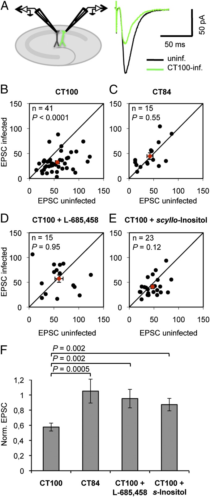
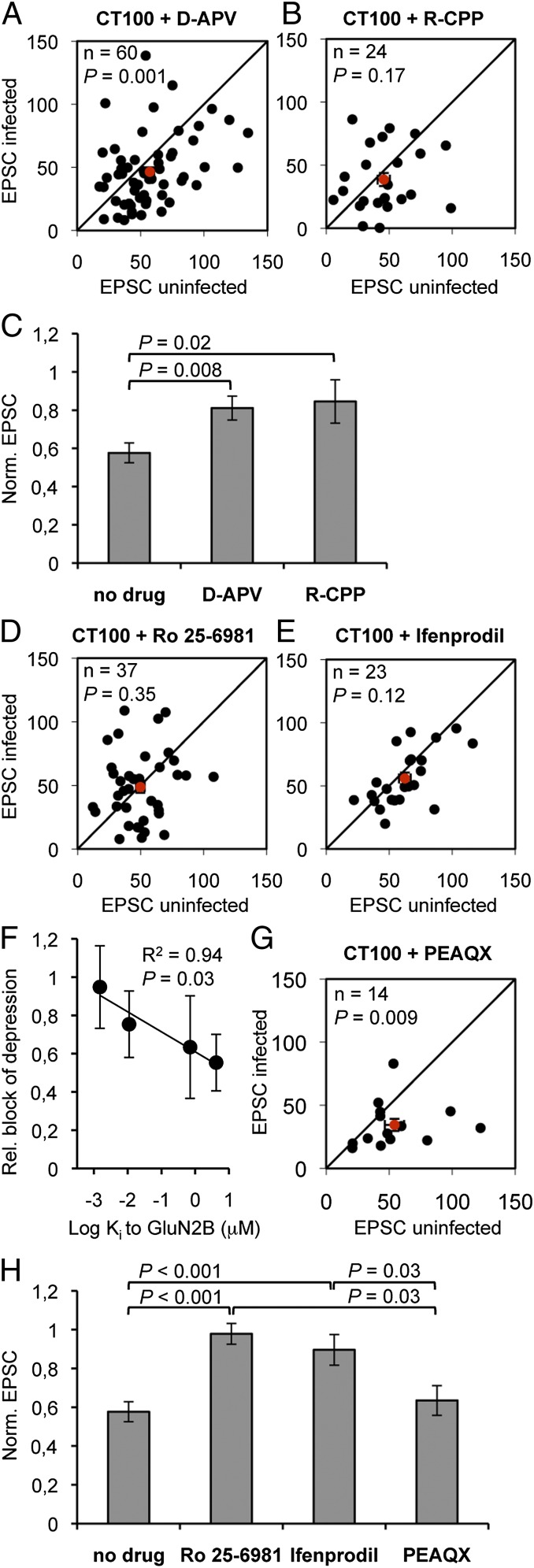
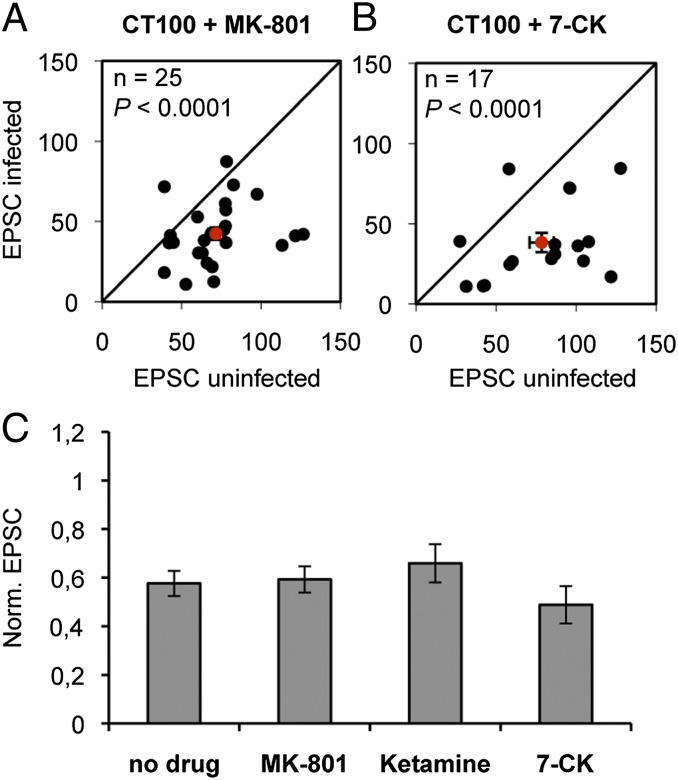
The mechanisms by which β-amyloid (Aβ), a peptide fragment believed to contribute to Alzheimer's disease, leads to synaptic deficits are not known. Here we find that elevated oligomeric Aβ requires ion flux-independent function of NMDA receptors (NMDARs) to produce synaptic depression. Aβ activates this metabotropic NMDAR function on GluN2B-containing NMDARs but not on those containing GluN2A. Furthermore, oligomeric Aβ leads to a selective loss of synaptic GluN2B responses, effecting a switch in subunit composition from GluN2B to GluN2A, a process normally observed during development. Our results suggest that conformational changes of the NMDAR, and not ion flow through its channel, are required for Aβ to produce synaptic depression and a switch in NMDAR composition. This Aβ-induced signaling mediated by alterations in GluN2B conformation may be a target for therapeutic intervention of Alzheimer's disease.


Conflict of interest statement

The authors declare no conflict of interest.

Figures

Fig. 1.
Fig. 1.
APP-CT100–expressing neurons display oligomeric Aβ-mediated synaptic AMPAR depression. (A) Model figure for a dual whole-cell recording of an infected and a neighboring uninfected CA1 neuron (Left) and an example trace of evoked AMPAR currents of such a recording (Right). (BE) Dot-plots of paired excitatory postsynaptic currents (EPSC) recordings, average (with error bars, SEM) denoted in red. (F) EPSCs of APP-CT100–infected neurons normalized to neighboring uninfected neurons upon incubation with indicated drugs. APP-CT100 (B), but not APP-CT84 expression (C) or incubation with the γ-secretase inhibitor L-685,458 (D), or incubation with 5 μM scyllo-Inositol (E), leads to synaptic AMPAR depression. Error bars, SEM. Statistics: paired Student t test of log-transformed data when comparing cell pairs (BE), nonpaired Student t test when comparing different conditions (F).
Fig. 2.
Fig. 2.
Oligmeric Aβ-mediated synaptic depression requires GluN2B-containing NMDAR function. (A, B, D, E, and G) Dot-plots of paired EPSC recordings, average denoted in red. (C and H) EPSCs of APP-CT100–infected neurons normalized to neighboring uninfected neurons upon incubation with indicated drugs. In slices incubated with 100 μM D-APV (A) or with 20 μM R-CPP (B) Aβ-driven synaptic AMPAR depression is significantly inhibited (C). Incubation with GluN2B-selective antagonists 3 μM Ro 25-6981 (D) or 30 μM ifenprodil (E) effectively blocked synaptic AMPAR depression in APP-CT100–expressing neurons. (F) For the four GluN2-specific drugs as shown in AE, their capacity to block the Aβ-mediated synaptic depression was plotted vs. their affinity (Ki) for GluN2B. (G) PEAQX (50 nM) did not block Aβ-driven synaptic AMPAR depression. (H) Bar graphs and statistical comparisons for data shown in dot-plots. Error bars, SEM. Statistics: paired Student t test of log-transformed data (A, B, D, E, and G), nonpaired Student t test (C and H), and regression analysis (F).
Fig. 3.
Fig. 3.
Oligmeric Aβ-mediated synaptic depression is independent of ion flux through the NMDAR. (A and B) Dot-plots of paired EPSC recordings, average denoted in red. (C) EPSCs of APP-CT100–infected neurons normalized to neighboring uninfected neurons upon incubation with indicated drugs. Incubation with 30 μM MK-801 (A and C), 200 μM ketamine (C), or 100 μM 7-Cl-kynurenate (B and C) did not block synaptic AMPAR depression in APP-CT100–expressing cells. Error bars, SEM. Statistics: paired Student t test of log-transformed data when comparing cell pairs, nonpaired Student t test when comparing different conditions.
Fig. 4.
Fig. 4.
Oligomeric Aβ selectively reduces GluN2B-mediated synaptic currents and increases the synaptic GluN2A to GluN2B ratio in an ion flux-independent manner. (A) Example traces of NMDAR currents from pairs of infected (green) and uninfected (black) CA1 neurons, before and after Ro 25-6981 wash-in to reveal GluN2A- and GluN2B-contributing NMDAR currents. (B) Paired NMDAR EPSCs of APP-CT100–infected neurons (green bars), normalized to neighboring uninfected neurons (white bars). (C) GluN2A to GluN2B current ratio of pairs of APP-CT100 infected and its neighboring uninfected CA1 neuron, individual pairs in gray, average in black. (A and B) APP-CT100 expression led to a selective loss in GluN2B currents and increase in GluN2A to GluN2B ratio. Both these effects were inhibited upon incubation of hippocampal slices with 100 μM D-APV during APP-CT100 expression but remained unaffected upon incubation with 200 μM ketamine (C). Error bars, SEM. Statistics: paired Student t test.

References

    1. Terry RD, et al. Physical basis of cognitive alterations in Alzheimer’s disease: Synapse loss is the major correlate of cognitive impairment. Ann Neurol. 1991;30(4):572–580. - PubMed
    1. Mucke L, et al. High-level neuronal expression of abeta 1-42 in wild-type human amyloid protein precursor transgenic mice: Synaptotoxicity without plaque formation. J Neurosci. 2000;20(11):4050–4058. - PMC - PubMed
    1. Shankar GM, et al. Amyloid-beta protein dimers isolated directly from Alzheimer’s brains impair synaptic plasticity and memory. Nat Med. 2008;14(8):837–842. - PMC - PubMed
    1. Lesné S, et al. A specific amyloid-beta protein assembly in the brain impairs memory. Nature. 2006;440(7082):352–357. - PubMed
    1. Cheng IH, et al. Accelerating amyloid-beta fibrillization reduces oligomer levels and functional deficits in Alzheimer disease mouse models. J Biol Chem. 2007;282(33):23818–23828. - PubMed

Publication types

MeSH terms