. 2013 Mar 5;110(10):4069-74.
doi: 10.1073/pnas.1218497110. Epub 2013 Feb 19.

Reactive astrocytes secrete lcn2 to promote neuron death

Affiliations

Reactive astrocytes secrete lcn2 to promote neuron death

Fangfang Bi et al. Proc Natl Acad Sci U S A. .

Abstract

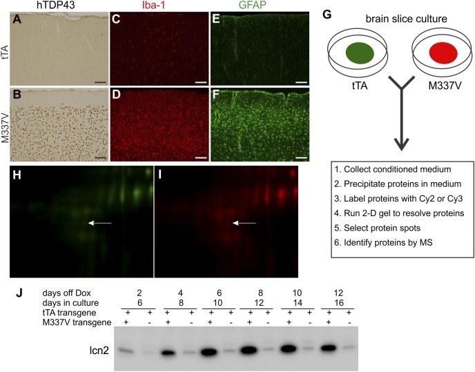
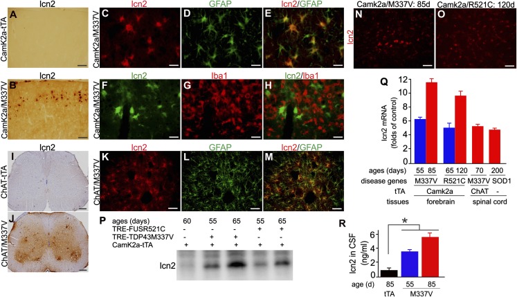
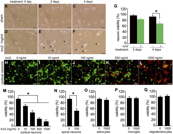
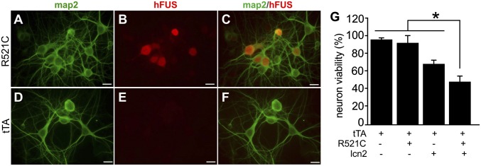
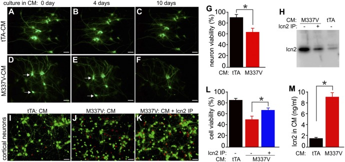
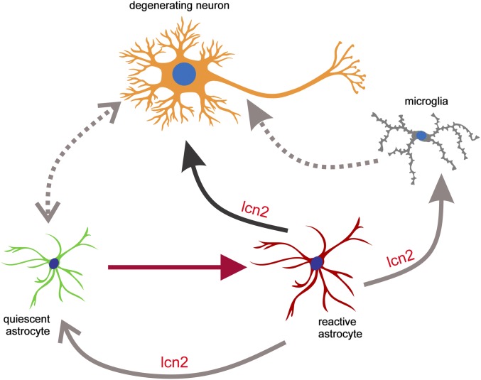
Glial reaction is a common feature of neurodegenerative diseases. Recent studies have suggested that reactive astrocytes gain neurotoxic properties, but exactly how reactive astrocytes contribute to neurotoxicity remains to be determined. Here, we identify lipocalin 2 (lcn2) as an inducible factor that is secreted by reactive astrocytes and that is selectively toxic to neurons. We show that lcn2 is induced in reactive astrocytes in transgenic rats with neuronal expression of mutant human TAR DNA-binding protein 43 (TDP-43) or RNA-binding protein fused in sarcoma (FUS). Therefore, lcn2 is induced in activated astrocytes in response to neurodegeneration, but its induction is independent of TDP-43 or FUS expression in astrocytes. We found that synthetic lcn2 is cytotoxic to primary neurons in a dose-dependent manner, but is innocuous to astrocytes, microglia, and oligodendrocytes. Lcn2 toxicity is increased in neurons that express a disease gene, such as mutant FUS or TDP-43. Conditioned medium from rat brain slice cultures with neuronal expression of mutant TDP-43 contains abundant lcn2 and is toxic to primary neurons as well as neurons in cultured brain slice from WT rats. Partial depletion of lcn2 by immunoprecipitation reduced conditioned medium-mediated neurotoxicity. Our data indicate that reactive astrocytes secrete lcn2, which is a potent neurotoxic mediator.


Conflict of interest statement

The authors declare no conflict of interest.

Figures

Fig. 1.
Fig. 1.
lcn2 is secreted from cultured brain slice expressing mutant human TDP-43. (A and B) Immunohistochemistry in rat cortex revealed that human TDP-43 with M337V substitution (hTDP43) was expressed in Camk2α-tTA/TRE-TDP43M337V double (B: M337V), but not Camk2α-tTA single (A: tTA), transgenic rats. (CF) Immunofluorescent staining demonstrated that microglia (C and D) and astrocytes (E and F) were substantially activated in M337V rats (D and F) compared with tTA rats (C and E). Rats were analyzed at the age of 55 d. (Scale bar: A–F, 100 µm.) (G) Schematic diagrams of the protocols for culturing brain slices and detecting secreted proteins in culture medium. (H and I) Representative images show that the fluorescent intensity of a protein spot corresponding to lcn2 was increased in the culture medium of a M337V brain slice (I, red) compared with a tTA brain slice culture (H, green). The entire 2D gel images are shown in Fig. S2. The brain slices were cultured for 4 d in the presence of Dox to allow for recovery from slicing and were then cultured in Dox-free medium to allow hTDP43 expression. After 8 days in vitro (DIV), brain slices were further cultured in serum-free medium for 24 h and the culture medium was collected for analysis of secreted proteins by 2D gels and MS. (J) Immunoblotting detected lcn2 in the culture medium. Each lane was loaded with 10 µL culture medium. Each well of a six-well plate was cultured with three slices from the same regions of tTA or M337V rat brains.
Fig. 2.
Fig. 2.
lcn2 is induced in reactive astrocytes in transgenic rats expressing mutant TDP-43, FUS, or SOD1. (A and B) Immunohistochemistry in rat cortex revealed significant lcn2 induction in Camk2α-tTA/TRE-TDP43M337V double (B, CamK2α/M337V), but not Camk2α-tTA single (A, CamK2α-tTA), transgenic rats. Rats were analyzed at 55 d of age. (C–H) Double-labeling fluorescent staining revealed that lcn2 (C and F) colocalized with GFAP (D and E) but not with Iba1 (G and H). (I and J) Immunohistochemistry revealed that lcn2 was substantially up-regulated in the spinal cord of ChAT-tTA/TRE-TDP43M337V double-transgenic rats (ChAT/M337V) compared with ChAT-tTA single-transgenic rats. (K–M) Immunofluorescence staining revealed that lcn2 colocalized with GFAP in ChAT/M337V rat spinal cord. ChAT-tTA and ChAT/M337V rats were analyzed at 58 d of age. (Scale bar: A and B, 100 µm; C–H, 30 µm; I and J, 200 µm; and K–M, 50 µm.) (N and O) Immunofluorescence staining revealed that lcn2 was diffusely expressed in mutant rat cortex at disease end-stages. (Scale bar: 30 µm.) (P) Immunoblotting revealed that lcn2 was up-regulated in rats expressing mutant TDP-43 or FUS. Rat frontal cortex was analyzed for lcn2 expression. (Q) Quantitative PCR revealed that lcn2 was up-regulated in rats expressing mutant TDP-43, FUS, or SOD1. Lcn2 mRNA was normalized to L17 mRNA and was calculated as a ratio of mutant rats to age-matched normal littermates. Data are mean ± SEM (n = 3–5). (R) ELISA lcn2 levels in the CSF of tTA and M337V transgenic rats. Data are mean ± SEM (n = 7), *P < 0.05.
Fig. 3.
Fig. 3.
lcn2 is selectively toxic to neurons. (A–F) Phase-contrast micrographs show that synthetic lcn2 induced progressive neuronal death. Cortical neurons were prepared from WT SD rat embryos and were cultured at low density for 10 d before lcn2 or sham treatment. (Scale bar: A–F, 50 µm.) (G) Cell counting revealed that lcn2 induced significant neuronal death. Cortical neurons were cultured in gridded chambers and examined daily under a microscope. The fates of chosen neurons were monitored throughout lcn2 treatment. Data are mean ± SEM (n = 6), *P < 0.05. (H–L) Micrographs show a dose-dependent response of cortical neurotoxicity to lcn2. Cortical neurons were grown at a high density and treated with varying doses of lcn2 for 4 d. Cell viability was examined with a cell Live/Dead assay. (Scale bar: H–L, 50 µm.) (M) Cell counting was performed to count the number of cortical neurons after lcn2 treatment. Data are mean ± SEM (n = 6), *P < 0.05. (N) Cell Live/Dead assay revealed the susceptibility of motor neurons to lcn2 toxicity. Mixed neurons were prepared from WT SD rat embryos and cultured for 10 d before they were treated with lcn2 for 4 d. Data are mean ± SEM (n = 7), *P < 0.05. (O–Q) Cell Live/Dead assay revealed the susceptibility of glial cells to lcn2 toxicity. Glial cells were prepared from WT SD rats (P3) and separated to enrich astrocytes, microglia, or oligodendrocytes. Enriched glial cells were treated with lcn2 for 4 d and analyzed for viability. Data are mean ± SEM (n = 5), *P < 0.05.
Fig. 4.
Fig. 4.
Expression of mutant FUS sensitizes cortical neurons to lcn2 toxicity. (A–F) Double immunofluorescent labeling revealed that human FUS with R521C substitution (hFUS) colocalized with the neuronal marker MAP2 in cortical neurons. hFUS expression was detected in the neurons of CamK2α-tTA/TRE-FUS-R521C double-transgenic rats (R521C) but not in the neurons of CamK2α-tTA single-transgenic rats (tTA). Enriched cortical neurons were prepared from rat embryos (E19) and cultured in the absence of Dox to allow for mutant hFUS expression. Neurons were examined for transgene expression after 10 d in culture. (Scale bar: A–F, 20 µm.) (G) Cell viability assay revealed the susceptibility of neurons to lcn2 toxicity. Enriched cortical neurons were cultured for 10 d and then treated with synthetic lcn2 (10 ng/mL) for 4 d before the viability assay. Data are mean ± SEM (n = 7), *P < 0.05.
Fig. 5.
Fig. 5.
lcn2 in CM from rat brain slice cultures is a potent mediator of neurotoxicity. (A–F) Representative images show that cortical neurons are sensitive to CM from brain slice cultures expressing mutant human TDP-43 (M337V-CM). Hippocampal slices (300 µm) were prepared from CamK2α-tTA/TRE-GFP double-transgenic rats (P1) that expressed GFP in neurons. Hippocampal slices were cultured in normal medium for 4 d before they were cultured in CM collected from brain slice cultures carrying CamK2α-tTA single (tTA) or Camk2α-tTA/TRE-TDP43M337V double (M337V) transgenes. Arrows indicate two GFP-labeled neurons that were lost by 10 d in culture. (Scale bar: A–F, 40 µm.) (G) Cell viability assay was used to quantify CM-induced cytotoxicity in rat brain slices expressing mutant human TDP-43. GFP-labeled neuron viability in hippocampal slices was determined after culture in CM for 10 d. Data are mean ± SEM (n = 5), *P < 0.05. (H) Immunoblotting revealed that lcn2 was partially depleted from the CM by immunoprecipitation (IP). CM from M337V slice cultures was immunoprecipitated with an lcn2 antibody, and the efficiency of lcn2 IP was examined by immunoblotting. Each lane was loaded with 10 µL CM. (I–K) Representative images illustrate the sensitivity of cortical neurons to CM with or without lcn2 depletion. Neurons were examined for viability after culture in CM for 10 d. (Scale bars: I–K, 40 µm.) (L) Cell Live/Dead assay revealed that CM-induced cytotoxicity was reduced after lcn2 immunoprecipitation. Data are mean ± SEM (n = 7), *P < 0.05. (M) ELISA revealed the levels of rat lcn2 in CM from tTA or M337V rat brain slice cultures. Data are mean ± SEM (n = 5), *P < 0.01.
Fig. 6.
Fig. 6.
Schematic diagram illustrating the hypothesized astrocytic secretion of lcn2 in response to neurodegeneration. Degenerating neurons activate quiescent astrocytes, and reactive astrocytes secrete lcn2 to promote neuronal death. Reactive astrocytes may also secrete lcn2 to promote astrocyte and microglia activation.

References

    1. Huang C, et al. Entorhinal cortical neurons are the primary targets of FUS mislocalization and ubiquitin aggregation in FUS transgenic rats. Hum Mol Genet. 2012;21(21):4602–4614. - PMC - PubMed
    1. Tong J, et al. XBP1 depletion precedes ubiquitin aggregation and Golgi fragmentation in TDP-43 transgenic rats. J Neurochem. 2012;123(3):406–416. - PMC - PubMed
    1. Escartin C, Bonvento G. Targeted activation of astrocytes: A potential neuroprotective strategy. Mol Neurobiol. 2008;38(3):231–241. - PubMed
    1. Lee S, et al. Lipocalin-2 is an autocrine mediator of reactive astrocytosis. J Neurosci. 2009;29(1):234–249. - PMC - PubMed
    1. Lee S, et al. A dual role of lipocalin 2 in the apoptosis and deramification of activated microglia. J Immunol. 2007;179(5):3231–3241. - PubMed

Publication types

MeSH terms