. 2013 Mar 19;110(12):4557-62.
doi: 10.1073/pnas.1222902110. Epub 2013 Feb 19.

Blastocyst complementation generates exogenic pancreas in vivo in apancreatic cloned pigs

Affiliations

Blastocyst complementation generates exogenic pancreas in vivo in apancreatic cloned pigs

Hitomi Matsunari et al. Proc Natl Acad Sci U S A. .

Abstract

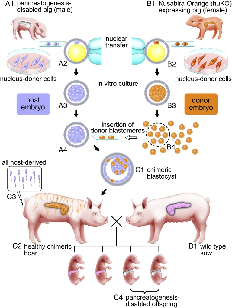
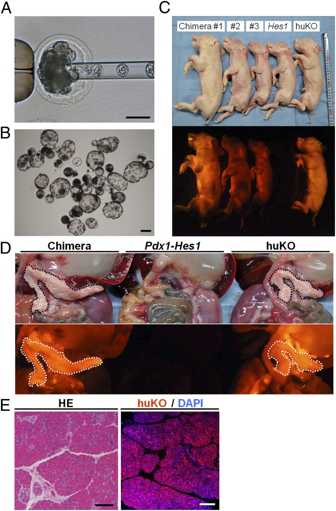
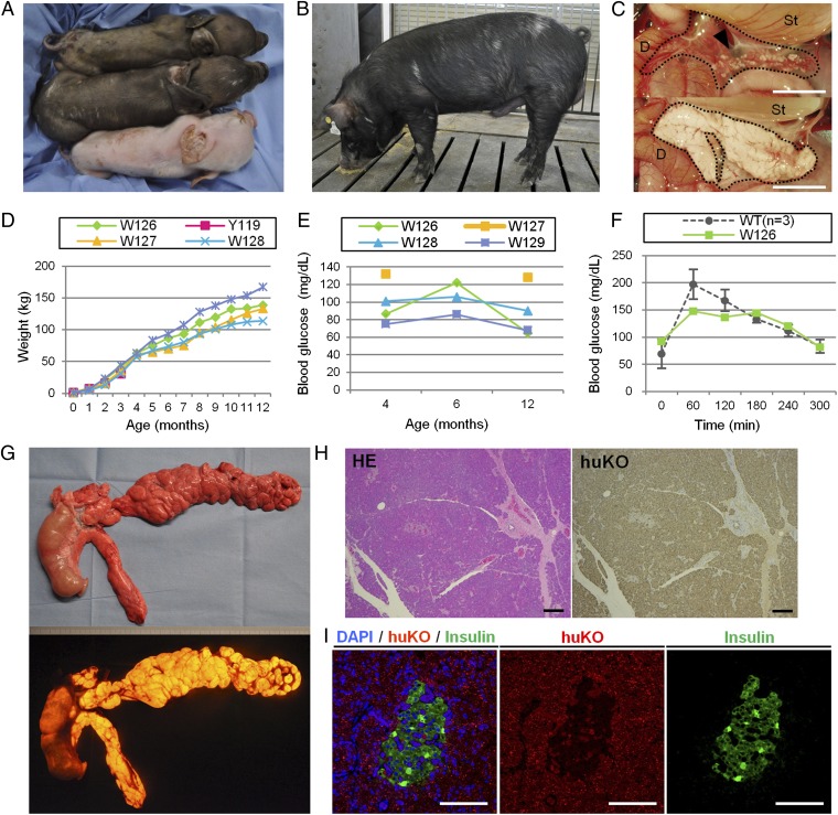
In the field of regenerative medicine, one of the ultimate goals is to generate functioning organs from pluripotent cells, such as ES cells or induced pluripotent stem cells (PSCs). We have recently generated functional pancreas and kidney from PSCs in pancreatogenesis- or nephrogenesis-disabled mice, providing proof of principle for organogenesis from PSCs in an embryo unable to form a specific organ. Key when applying the principles of in vivo generation to human organs is compensation for an empty developmental niche in large nonrodent mammals. Here, we show that the blastocyst complementation system can be applied in the pig using somatic cell cloning technology. Transgenic approaches permitted generation of porcine somatic cell cloned embryos with an apancreatic phenotype. Complementation of these embryos with allogenic blastomeres then created functioning pancreata in the vacant niches. These results clearly indicate that a missing organ can be generated from exogenous cells when functionally normal pluripotent cells chimerize a cloned dysorganogenetic embryo. The feasibility of blastocyst complementation using cloned porcine embryos allows experimentation toward the in vivo generation of functional organs from xenogenic PSCs in large animals.


Conflict of interest statement

The authors declare no conflict of interest.

Figures

Fig. 1.
Fig. 1.
Generation of pancreatogenesis-disabled pigs. (A) Construction of a Pdx1-Hes1 expression vector consisting of the mouse Pdx1 promoter, mouse Hes1 cDNA, and rabbit β-globin 3′ flanking sequence, including the polyadenylation signal (pA). (B and C) Macroscopic and microscopic appearances of the vestigial pancreas (arrowhead) of a Pdx1-Hes1 transgenic (Tg) male fetus and the pancreas of a WT fetus at the same gestational age. (D, Upper) Faithful reproduction of the pancreatogenesis-disabled phenotype of the Pdx1-Hes1 Tg fetus in fetuses cloned from it. (D, Lower) Normal pancreata of age-matched WT fetuses. D, duodenum; Ki, kidney; Sp, spleen; St, stomach. (Scale bars: B and D, 5 mm; C, 50 μm.)
Fig. 2.
Fig. 2.
Schematic representation of complementation for Pdx1-Hes1 cloned pig embryos with a pancreatogenesis-disabled phenotype using cloned embryos expressing huKO. Primary fibroblast cells as nucleus donor cells for somatic cell cloning were established from a pancreatogenesis-disabled cloned pig with Pdx1-Hes1 transgene expression (A1) and a cloned pig with systemic orange fluorescence conferred by huKO transgene expression (B1). (A2A4) Host embryos reconstructed by nuclear transfer from male Pdx1-Hes1 transgenic cells yielded pancreatogenesis-disabled piglets. (B2 and B3) Donor embryos were reconstructed by nuclear transfer from female cells expressing huKO. Blastomeres isolated from donor embryos at the morula stage (B4) were inserted into host embryo morulae (A4) to produce chimeric blastocysts (C1) and pigs (C2). (C2) All the chimeric pigs obtained developed into fertile males as a result of intersex chimerism between the male host embryos and female donor embryos. (C3) Sperm of the chimeric boars theoretically originate from male host embryos carrying the Pdx1-Hes1 transgene. After mating of chimeric boars with WT sows (D1), the pancreatogenesis-disabled phenotype of the Pdx1-Hes1 host embryos was transmitted to the next generation (C4).
Fig. 3.
Fig. 3.
Construction of chimeric embryos and fetuses from Pdx1-Hes1 cloned and huKO cloned embryos. (A) Cloned embryo derived from the Pdx1-Hes1 transgenic fetus via microinjection with donor morula blastomeres. (B) Chimeric blastocysts. (C) Full-term chimeric fetuses (chimeras 1–3) and sibling nonchimeric cloned fetuses derived from host (Hes1) or donor (huKO) embryos. Note that nonchimeric fetuses derived from host embryos (Hes1) showed no fluorescence. (D, Center) Fetuses derived from host embryos were pancreatogenesis-disabled. Pancreata of the chimeric fetuses (Left) appeared normal and brightly fluoresced orange throughout, as did pancreata of huKO clone fetuses (Right), indicating that pancreata of the chimeric fetuses were generated from donor embryo cells. (E) Almost all pancreatic tissue of chimeric fetuses stained with anti-huKO antibody. HE, H&E. (Scale bars: A, B, and E, 100 μm.)
Fig. 4.
Fig. 4.
Production of chimeric offspring by complementation of Pdx1-Hes1 pancreatogenesis-disabled embryos. (A) Chimeric pig (middle pig) was obtained after complementation of host Pdx1-Hes1 embryos with embryonic cells from a coat-colored WT donor. Sibling nonchimeric cloned pigs were derived from donor (top pig, brown coat) and host (bottom pig, white coat) embryos. (B) Mature chimeric boar exhibits WT (donor) coat-color chimerism. (C) Vestigial pancreas (Upper, arrowhead) of a host embryo-derived cloned piglet and normal pancreas (Lower) of a donor-embryo derived piglet. D, duodenum; St, stomach. (D and E) Normal growth and serum glucose concentrations were observed in chimeric pigs. Pig W127 did not undergo blood sampling at the age of 6 mo due to a leg injury. (F) Serum glucose concentrations in a chimeric pig (W126) and WT pig during oral glucose tolerance testing. (G) Normally formed pancreas generated in a chimeric pig (W128) exhibits orange fluorescence derived from the donor huKO embryo. (H) Histological appearance of pancreas generated in a chimeric pig, with staining throughout by anti-huKO antibody. HE, H&E. (I) Islet of Langerhans in the pancreas generated in a chimeric pig marks immunohistochemically for insulin. (Scale bars: C, 1 cm; H and I, 100 μm.)

Comment in

References

    1. Lindvall O, Kokaia Z. Stem cells for the treatment of neurological disorders. Nature. 2006;441(7097):1094–1096. - PubMed
    1. MacLaren RE, et al. Retinal repair by transplantation of photoreceptor precursors. Nature. 2006;444(7116):203–207. - PubMed
    1. Kobayashi T, et al. Generation of rat pancreas in mouse by interspecific blastocyst injection of pluripotent stem cells. Cell. 2010;142(5):787–799. - PubMed
    1. Usui JI, et al. Generation of kidney from pluripotent stem cells via blastocyst complementation. Am J Pathol. 2012;180(6):2417–2426. - PubMed
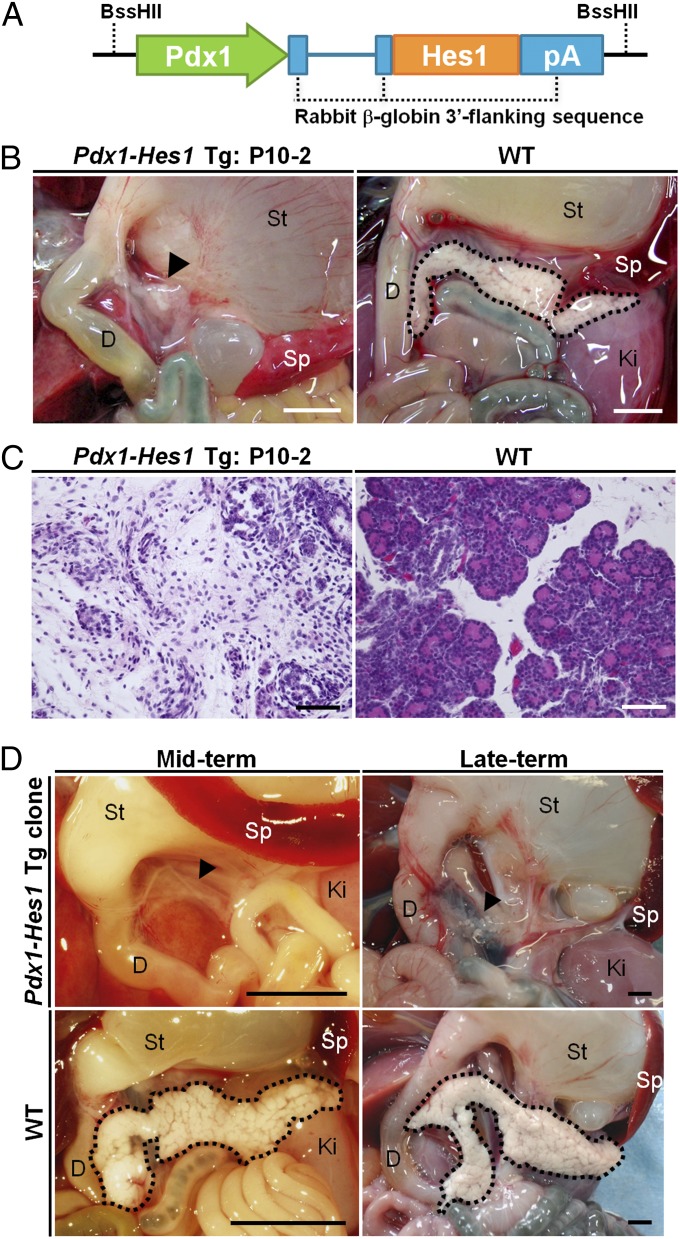
    1. Sumazaki R, et al. Conversion of biliary system to pancreatic tissue in Hes1-deficient mice. Nat Genet. 2004;36(1):83–87. - PubMed

Publication types