

doi: 10.1073/pnas.1216691110.
Epub 2013 Feb 19.
Structural basis for potent inhibitory activity of the antibiotic tigecycline during protein synthesis
Affiliations
- PMID: 23431179
- PMCID: PMC3593886
- DOI: 10.1073/pnas.1216691110
Item in Clipboard
Structural basis for potent inhibitory activity of the antibiotic tigecycline during protein synthesis
Proc Natl Acad Sci U S A.
.
Abstract
Here we present an X-ray crystallography structure of the clinically relevant tigecycline antibiotic bound to the 70S ribosome. Our structural and biochemical analysis indicate that the enhanced potency of tigecycline results from a stacking interaction with nucleobase C1054 within the decoding site of the ribosome. Single-molecule fluorescence resonance energy transfer studies reveal that, during decoding, tigecycline inhibits the initial codon recognition step of tRNA accommodation and prevents rescue by the tetracycline-resistance protein TetM.
Conflict of interest statement
The authors declare no conflict of interest.
Figures
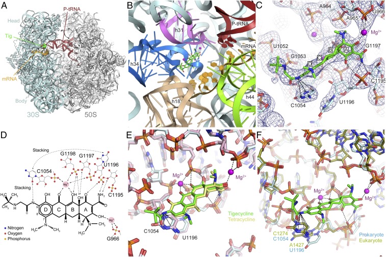
X-ray crystal structure of tigecycline on the 70S ribosome. (A) Overview from the A site of the 70S ribosome with tRNAfMet in the P site (red), mRNA (orange) and tigecycline (green) bound. (B) View of the tigecycline binding site showing the rRNA elements in the vicinity of the site. (C) The fully refined electron density map (2Fobs−Fcalc) contoured at 1.2 sigma for the area surrounding the tigecycline binding site. Dashed lines indicate the stacking of the 9-t-butylglycylamido moiety of tigecycline with nucleobase C1054 and the coordination of the additional Mg2+ connecting tigecycline to G966 (h31). (D) Schematic chemical structure of tigecycline showing possible hydrogen bonds and other interactions with Mg2+ ions and bases from 16S rRNA. (E) Comparison of the binding modes of tigecycline (green) and tetracycline (yellow) via superimposition of the 16S rRNA. (F) Comparison of the prokaryotic T. thermophilus and the eukaryotic Saccharomyces cerevisiae tetracycline binding sites by superimposition of h34. Note that the nucleotide equivalents to C1054 and U1196 in S. cerevisiae are C1274 and A1427, the latter of which is slightly shifted, whereas the rest of the binding pocket is nearly identical.
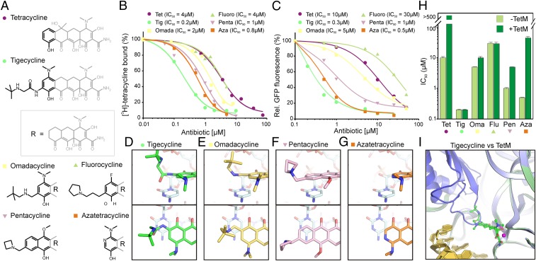
Binding and inhibitory properties of tetracycline derivatives. (A) Chemical structures of tetracycline, tigecycline, omadacycline, 9-propylpyrrolidyl-7-fluorocyline (fluorocycline), 7-methoxy-10-azetidinomethyl pentacycline (pentacycline), and 7-dimethylamido 8-azatetracycline (azatetracycline). (B) The inhibitory effect of tetracycline derivatives from A were monitored by using an E. coli in vitro transcription/translation assay monitoring the fluorescence of GFP as a function of antibiotic concentration. (C) The ability of tetracycline compounds from panel A to compete for binding to the E. coli 70S ribosome with [3H]tetracycline was monitored as a function of antibiotic concentration. (D–G) The stacking interaction of the (D) glycyl side chain of tigecycline (green) as observed in the tigecycline⋅70S structure is compared with models for the (E) omadacycline (yellow), (F) pentacycline (pink), and (G) azatetracycline (orange) docked on the 70S ribosome based on the tigecycline⋅70S structure. (H) The ability of the tetracycline derivatives from panel A to overcome TetM-mediated resistance was determined by performing translation inhibition assays as in C in the absence (light green) and presence (dark green) of TetM. The IC50 is presented as a log-scale (in µM). (I) Superimposition of the 70S ribosome structure with TetM (19) (blue) on the 70S structure with tigecycline (green) and mRNA (11) (gold).
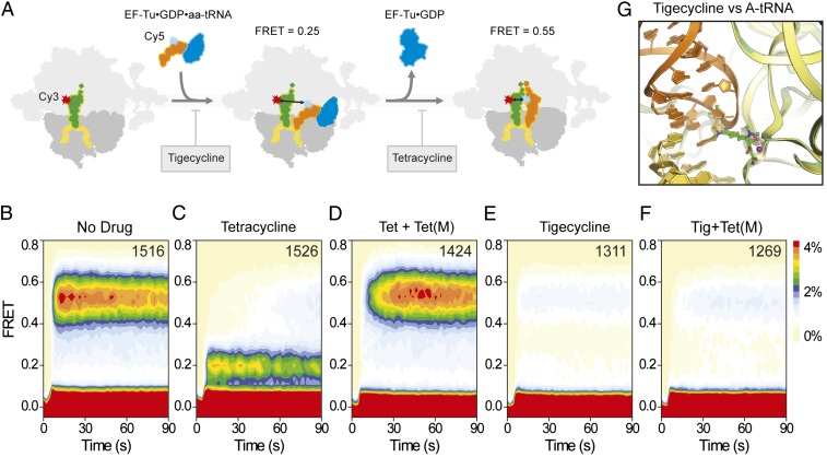
Effect of tetracyclines and glycylcyclines on tRNA selection. (A) Schematic illustrating the delivery of EF-Tu⋅tRNA⋅GTP ternary complex containing cognate Phe-tRNAPhe(Cy5-acp3U47) to 70S E. coli ribosomes containing P-site OH-tRNAfMet(Cy3-s4U8), leading to low (0.2) FRET during initial steps of codon recognition, and high (0.55) FRET upon A-site tRNA accommodation. Tigecycline is more effective than tetracycline at blocking the initial selection process. Tetracycline is nevertheless effective at preventing transitions into the fully accommodated, high-FRET state. (B–F) Single-molecule FRET imaging of aa-tRNA selection performed under direct 532-nm excitation following a 5-min incubation (B) in the absence of drugs or with (C) 40 µM tetracycline, (D) 40 µM tetracycline and 0.1 µM TetM, (E) 2 µM tigecycline, or (F) 2 µM tigecycline and 0.1 µM TetM.
References
-
- Wilson DN. The A-Z of bacterial translation inhibitors. Crit Rev Biochem Mol Biol. 2009;44(6):393–433. - PubMed
-
- Brodersen DE, et al. The structural basis for the action of the antibiotics tetracycline, pactamycin, and hygromycin B on the 30S ribosomal subunit. Cell. 2000;103(7):1143–1154. - PubMed
-
- Murray JB, et al. Interactions of designer antibiotics and the bacterial ribosomal aminoacyl-tRNA site. Chem Biol. 2006;13(2):129–138. - PubMed
Publication types
MeSH terms
Substances
Associated data
- Actions
- Actions
- Actions
- Actions
Grants and funding
LinkOut - more resources
Full Text Sources
Other Literature Sources
Medical
Molecular Biology Databases
