. 2013 Jan;9(1):e1003120.
doi: 10.1371/journal.ppat.1003120. Epub 2013 Jan 31.

Discovery of a siderophore export system essential for virulence of Mycobacterium tuberculosis

Affiliations

Discovery of a siderophore export system essential for virulence of Mycobacterium tuberculosis

Ryan M Wells et al. PLoS Pathog. 2013 Jan.

Abstract

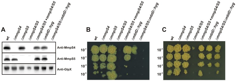
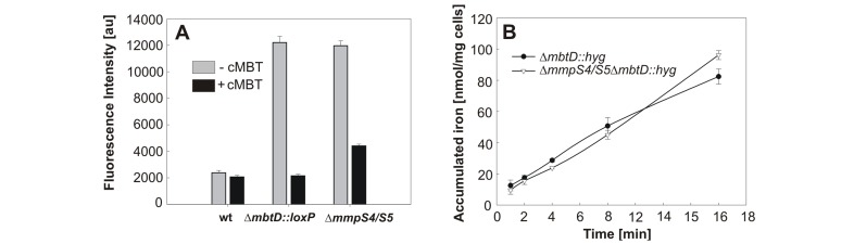
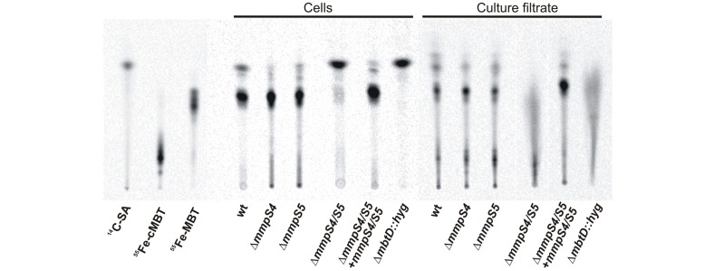
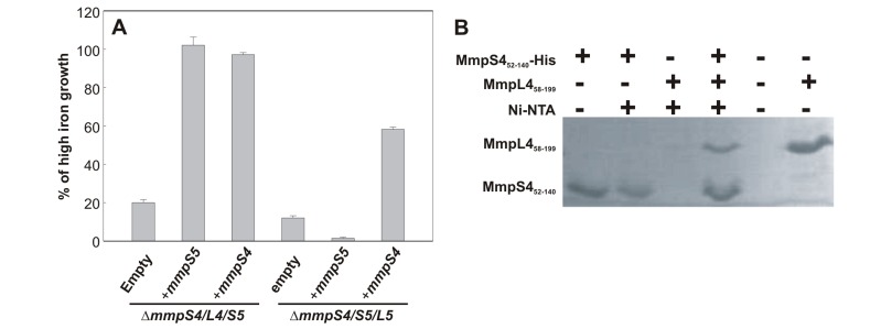
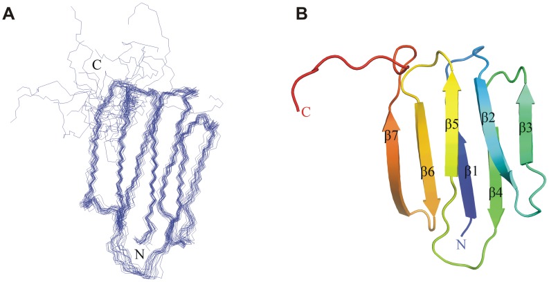
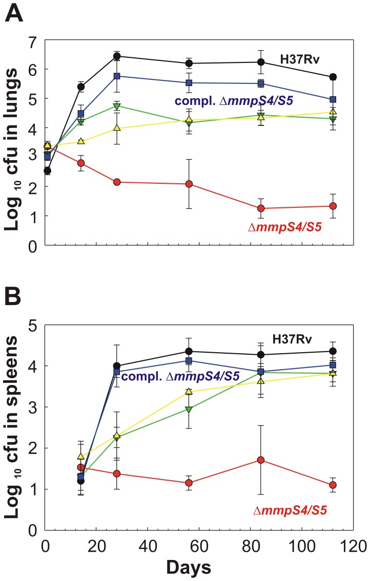
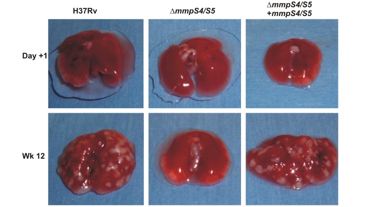
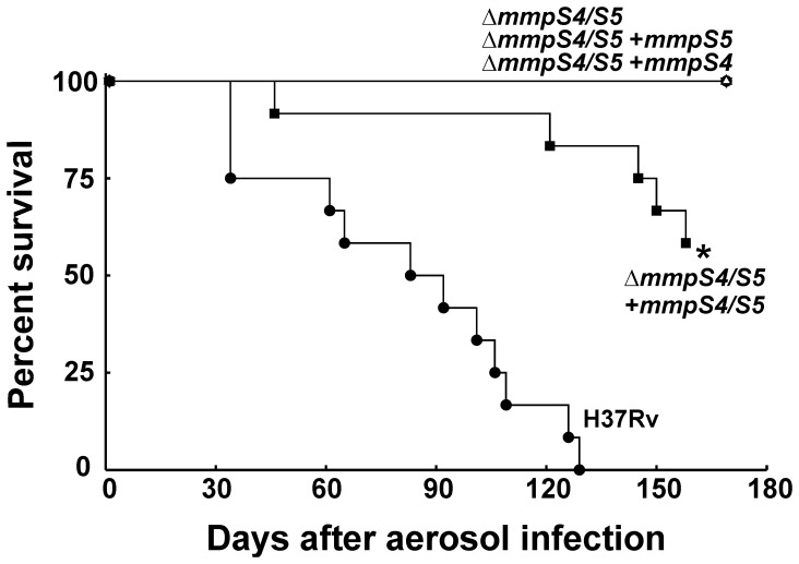
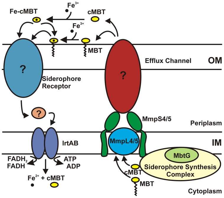
Iron is an essential nutrient for most bacterial pathogens, but is restricted by the host immune system. Mycobacterium tuberculosis (Mtb) utilizes two classes of small molecules, mycobactins and carboxymycobactins, to capture iron from the human host. Here, we show that an Mtb mutant lacking the mmpS4 and mmpS5 genes did not grow under low iron conditions. A cytoplasmic iron reporter indicated that the double mutant experienced iron starvation even under high-iron conditions. Loss of mmpS4 and mmpS5 did not change uptake of carboxymycobactin by Mtb. Thin layer chromatography showed that the ΔmmpS4/S5 mutant was strongly impaired in biosynthesis and secretion of siderophores. Pull-down experiments with purified proteins demonstrated that MmpS4 binds to a periplasmic loop of the associated transporter protein MmpL4. This interaction was corroborated by genetic experiments. While MmpS5 interacted only with MmpL5, MmpS4 interacted with both MmpL4 and MmpL5. These results identified MmpS4/MmpL4 and MmpS5/MmpL5 as siderophore export systems in Mtb and revealed that the MmpL proteins transport small molecules other than lipids. MmpS4 and MmpS5 resemble periplasmic adapter proteins of tripartite efflux pumps of Gram-negative bacteria, however, they are not only required for export but also for efficient siderophore synthesis. Membrane association of MbtG suggests a link between siderophore synthesis and transport. The structure of the soluble domain of MmpS4 (residues 52-140) was solved by NMR and indicates that mycobacterial MmpS proteins constitute a novel class of transport accessory proteins. The bacterial burden of the mmpS4/S5 deletion mutant in mouse lungs was lower by 10,000-fold and none of the infected mice died within 180 days compared to wild-type Mtb. This is the strongest attenuation observed so far for Mtb mutants lacking genes involved in iron utilization. In conclusion, this study identified the first components of novel siderophore export systems which are essential for virulence of Mtb.


Conflict of interest statement

The authors have declared that no competing interests exist.

Figures

Figure 1
Figure 1. MmpS4 or MmpS5 is required for growth of M. tuberculosis under iron-limited conditions.
A. Expression of MmpS4 and MmpS5 in Mtb. Whole cell lysates from wt Mtb mc26230, ΔmmpS4, ΔmmpS5, ΔmmpS4/S5, ΔmmpS4/S5+mmpS4/S5, ΔmbtD::hyg, and ΔmmpS4/S5/mbtD::hyg were probed by Western blot by using rabbit polyclonal antibodies raised against MmpS4 and MmpS5. The cytoplasmic fructose 1,6-bisphosphatase GlpX was used as a loading control and detected using an anti-GlpX antiserum. B, C. Serial dilutions of log-phase cultures of Mtb mc26230, ΔmmpS4, ΔmmpS5, ΔmmpS4/S5, ΔmmpS4/S5 fully complemented with mmpS4 and mmpS5, ΔmbtD::hyg, and ΔmmpS4/S5ΔmbtD::hyg were spotted on low iron glycerol-alanine-salts (GAS) plates (B), or on low iron GAS plates with 5 µM hemoglobin as an iron source (C).
Figure 2
Figure 2. MmpS4 and MmpS5 are not involved in iron sensing or uptake of siderophores.
A. GFP fluorescence was measured in wt Mtb mc26230, ΔmmpS4/S5, and ΔmbtD::loxP strains containing a gfp-based iron-regulated reporter construct. Strains were grown in 7H9 media and fluorescence was measured two days after the addition of carboxymycobactin (cMBT) (black bars) or blank control (grey bars). Experiments were performed in triplicate and are shown with standard deviations. B. Uptake of 55Fe loaded cMBT by Mtb ΔmmpS4/S5 (black circles) and ΔmmpS4/S5 ΔmbtD::hyg (white triangles). Assays were performed at 37°C using a final concentration of 0.25 µM cMBT, 0.45 µCi 55Fe in triplicate. Standard deviations are shown.
Figure 3
Figure 3. MmpS4 and MmpS5 are required for siderophore secretion in M. tuberculosis.
TLC of cell-associated and secreted siderophores extracted from cultures of wt Mtb H37Rv parent strain ML617, ΔmmpS4 single deletion mutant ML472, ΔmmpS5 single deletion mutant ML405, mmpS4/S5 double deletion mutant ML618, ΔmmpS4/S5 double deletion mutant fully complemented with mmpS4 and mmpS5 ML624, and the siderophore biosynthetic mutant ΔmbtD::hyg ML1424. Cultures were labelled with 7-[14C]-salicylic acid, which was run on the TLC as a control alongside 55Fe-loaded cMBT and mycobactin (MBT). Lanes containing cell-associated extracts were loaded with 5,000 cpm, while media extracts were loaded with 7,500 cpm.
Figure 4
Figure 4. MmpS4, MmpS5 and MbtG are membrane-associated proteins.
Proteins of subcellular fractions of wt Mtb (ML878) were extracted with 3% SDS and analyzed by SDS-polyacrylamide (10%) gel electrophoresis and Western blot using protein specific antibodies. A. Subcellular localization of MmpS4 and MmpS5. OmpATb, IdeR and Ag85 were used as controls for membrane, water-soluble cytoplasmic or periplasmic proteins and secreted proteins, respectively. MmpS4 and MmpS5 were detected using rabbit polyclonal antibodies. B. Subcellular localization of MbtG. MctB and IdeR were used as controls for membrane and water-soluble cytoplasmic or periplasmic proteins, respectively. MbtG was expressed with a C-terminal fusion of the Human influenza hemagglutinin (HA) tag which was detected using an HA-specific antibody.
Figure 5
Figure 5. MmpS proteins interact with MmpL proteins.
A. Genetic interactions between MmpS4 and MmpS5 proteins and their cognate MmpL proteins. Percent of growth in iron-restricted medium (7H9 medium containing 50 µM 2,2′-dipyridyl) of triple mutants ΔmmpS4/L4/S5 and ΔmmpS4/S5/L5 strains and those strains complemented with mmpS4 or mmpS5 compared to growth in iron-rich media. B. Interaction of the C-terminal soluble domain of MmpS4 (residues 52–140) with the L1 loop of MmpL4 (residues 58–199) by an in vitro pull down assay.
Figure 6
Figure 6. Structure of the C-terminal soluble domain of MmpS4 (residues 52–140).
A. NMR structure of MmpS452–140 showing the backbone superposition of the final 20 conformers. The coordinates for the structures have been deposited in the Protein Data Bank (PDB accession code 2LW3). B. Cartoon depiction of a representative structure.
Figure 7
Figure 7. MmpS4 and MmpS5 are required for virulence of M. tuberculosis in mice.
Colony forming unit (CFU) counts in lungs (A) and spleens (B) of mice infected with either the wt Mtb H37Rv parent strain ML617 (brown circles), the ΔmmpS4/S5 double deletion mutant ML618 (orange circles), the ΔmmpS4/S5 mutant singly complemented with mmpS5 ML619 (olive upside down triangles), the ΔmmpS4/S5 mutant singly complemented with mmpS4 ML620 (green triangles), or the ΔmmpS4/S5 mutant fully complemented with both mmpS4 and mmpS5 ML624 (aqua squares). Each data point represents the average of CFUs from the organs of four mice with standard deviations shown.
Figure 8
Figure 8. Gross pathology of mouse lungs infected with M. tuberculosis.
Gross pathology of whole lungs of BALB/c mice infected with wt Mtb H37Rv (ML617), ΔmmpS4/S5 (ML618), and ΔmmpS4/S5 complemented with both mmpS4 and mmpS5 (ML624).
Figure 9
Figure 9. Effect of mmpS4 and mmpS5 on the survival of mice infected with M. tuberculosis.
Survival of mice infected with wt Mtb H37Rv (ML617), ΔmmpS4/S5 (ML618), ΔmmpS4/S5 singly complemented with mmpS5 (ML619), ΔmmpS4/S5 singly complemented with mmpS4 (ML620), or ΔmmpS4/S5 fully complemented with mmpS4 and mmpS5 (ML624). Thirteen mice were infected with each strain. Mice were euthanized at day 169.
Figure 10
Figure 10. Model of siderophore–mediated iron uptake by M. tuberculosis.
Siderophores are synthesized by cytoplasmic synthases that function as a complex. Synthesis and transport of siderophores is likely coupled and dependent on the activity of the RND transporters, MmpL4 and MmpL5. Siderophore export requires that the membrane-associated proteins MmpS4 and MmpS5 function together with their cognate MmpL proteins. MmpS4 and MmpS5 are anchored to the inner membrane and may function as periplasmic adaptor proteins. Export of siderophores across the OM would require a yet undiscovered OMP. Once secreted, siderophores bind iron and would require an OMP siderophore receptor for transport across the OM. Once in the periplasmic space IrtAB imports ferric-siderophores across the inner membrane where iron is released from the siderophore and becomes available for the cell.

References

    1. Skaar EP (2010) The battle for iron between bacterial pathogens and their vertebrate hosts. PLoS Pathog 6: e1000949. - PMC - PubMed
    1. Searle S, Bright NA, Roach TI, Atkinson PG, Barton CH, et al. (1998) Localisation of Nramp1 in macrophages: modulation with activation and infection. J Cell Sci 111: 2855–2866. - PubMed
    1. Cellier MF, Courville P, Campion C (2007) Nramp1 phagocyte intracellular metal withdrawal defense. Microbes Infect 9: 1662–1670. - PubMed
    1. Wagner D, Maser J, Lai B, Cai Z, Barry CE 3rd, et al. (2005) Elemental analysis of Mycobacterium avium-, Mycobacterium tuberculosis-, and Mycobacterium smegmatis-containing phagosomes indicates pathogen-induced microenvironments within the host cell's endosomal system. J Immunol 174: 1491–1500. - PubMed
    1. de Voss JJ, Rutter K, Schroeder BG, Su H, Zhu Y, et al. (2000) The salicylate-derived mycobactin siderophores of Mycobacterium tuberculosis are essential for growth in macrophages. Proc Natl Acad Sci USA 97: 1252–1257. - PMC - PubMed

Publication types

MeSH terms