. 2013:2013:329606.
doi: 10.1155/2013/329606. Epub 2013 Feb 3.

Insulin resistance and muscle metabolism in chronic kidney disease

Affiliations

Insulin resistance and muscle metabolism in chronic kidney disease

James L Bailey. ISRN Endocrinol. 2013.

Abstract

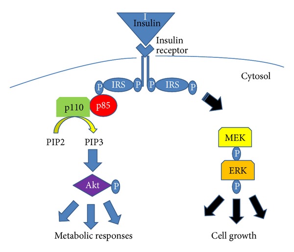
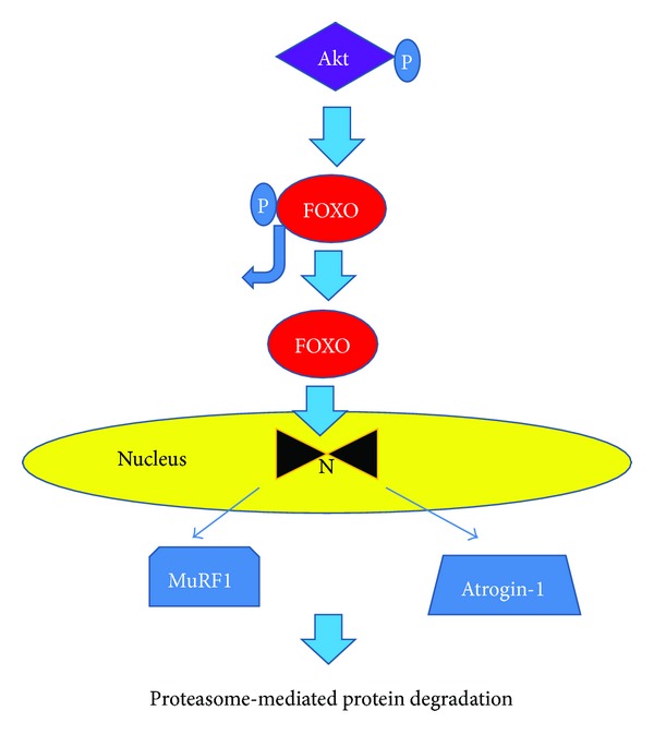
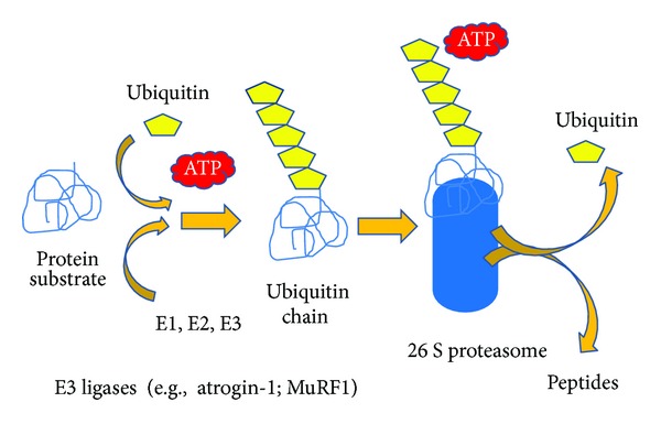
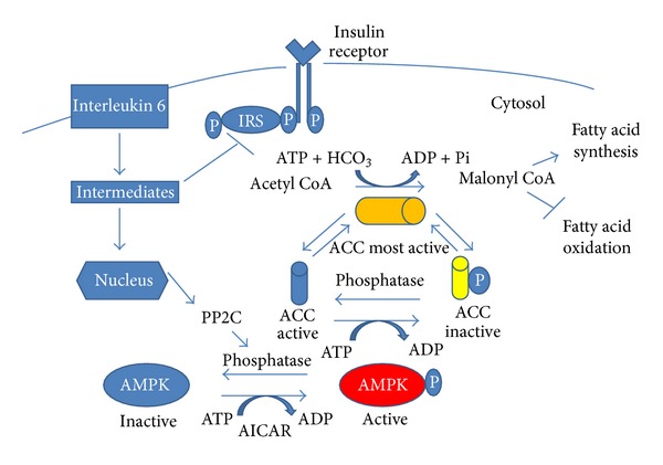
Insulin resistance is a common finding in chronic kidney disease (CKD) and is manifested by mild fasting hyperglycemia and abnormal glucose tolerance testing. Circulating levels of glucocorticoids are high. In muscle, changes in the insulin signaling pathway occur. An increase in the regulatory p85 subunit of Class I phosphatidylinositol 3-Kinase enzyme leads to decreased activation of the downstream effector protein kinase B (Akt). Mechanisms promoting muscle proteolysis and atrophy are unleashed. The link of Akt to the ubiquitin proteasome pathway, a major degradation pathway in muscle, is discussed. Another factor associated with insulin resistance in CKD is angiotensin II (Ang II) which appears to induce its intracellular effects through inflammatory cytokines or reactive oxygen species. Skeletal muscle ATP is depleted and the ability of AMP-activated protein kinase (AMPK) to replenish energy stores is blocked. How this can be reversed is discussed. Interleukin-6 (IL-6) levels are elevated in CKD and impair insulin signaling at the level of IRS-1. With exercise, IL-6 levels are reduced; glucose uptake and utilization are increased. For patients with CKD, exercise may improve insulin signaling and build up muscle. Treatment strategies for preventing muscle atrophy are discussed.


Figures

Figure 1
Figure 1
Insulin or insulin-like growth factor 1 (IGF-1) binds to its receptor and activates the receptor tyrosine kinase. The receptor undergoes autophosphorylation and provides a binding site for the insulin receptor substrate (IRS) proteins. Once bound, these IRS proteins undergo phosphorylation on tyrosine residues. These phosphorylated tyrosine residues provide a docking site for the p85 regulatory subunit of the Class I phosphatidylinositol 3-kinase. In turn, the p110 catalytic subunit is released, becomes activated and catalyzes the production of phosphatidylinositol (3, 4, 5)-triphosphate (PIP3) from phosphatidylinositol (3, 4)-biphosphate (PIP2). PIP3 then activates protein kinase B (Akt). Akt then serves as a branch point for a variety of downstream signaling pathways. Insulin and IGF-1 can also stimulate cell growth through the mitogen activated protein kinase pathway/extracellular signal related kinase (MEK/ERK) pathway.
Figure 2
Figure 2
A downstream target of protein Kinase B (Akt) is the Forkhead box O or FOXO transcription factors. Phosphorylation of the FOXOs by Akt deactivates them and prevents them from translocating to the nucleus. Dephosphorylated FOXOs translocate to the nucleus where they increase the expression of a variety of genes that suppress skeletal muscle hypertrophy and result in muscle atrophy. They also induce ubiquitin ligases such as muscle ring finger-1 (MuRF1) and atrogin-1 that promote skeletal muscle proteolysis.
Figure 3
Figure 3
A protein designated for catabolism is bound to a series of ubiquitin (Ub) molecules in a process requiring ATP. Initially, free Ub is bound to the Ub-activating enzyme E1 in an ATP dependent process. Ub is subsequently shuttled from the Ub-activating enzyme E1 to Ub-conjugating enzyme E2 through the formation of a thioester bond between Ub and a cysteine residue of the E2 enzyme. The Ub monomer is then conjugated to the target protein through a peptide bond between the ε-amino group of a lysine residue in the target protein and the carboxy-terminal glycine residue in Ub via the action of a Ub-ligase enzyme E3. At least 4 Ub monomers must be attached to the protein before the target protein can be recognized and degraded by the 26S proteasome. In the degradation process, peptides are formed and the ubiquitin is released where it can be recycled again.
Figure 4
Figure 4
Infusion of angiotensin II (Ang II) is associated with an upregulation of various cytokines including interleukin 6 (IL-6), tumor necrosis factor (TNF), and reactive oxygen species. Chronic exposure to IL-6 impairs insulin signaling at the level of insulin receptor substrate (IRS-1) through mechanisms that involve activation of proinflammatory kinases. There is also upregulation of protein phosphatase 2Ca (PP2Ca) which is known to dephosphorylate and inactivate AMP-activated protein kinase (AMPK). Downstream targets of AMPK signaling including peroxisome-proliferator-activated receptor gamma coactivator-1α (PGC1-α) and acetyl-coenzyme A carboxylase (ACC) are reduced. When ACC is phosphorylated, it is inactive and no longer able to catalyze the synthesis of malonyl-coenzyme A. Fatty acid oxidation is blocked and muscle ATP depletion occurs.
Figure 5
Figure 5
The precursor of myostatin is promyostatin and consists of a propeptide that binds to myostatin noncovalently to form an inactive complex. When myostatin is activated, it binds to its receptor; activin A bound to the extra cellular domain of a type II receptor (ActRIIB), which is present on muscle membranes. In turn ALK4 or ALK5 phosphorylates intracellular proteins called Smad2/3 and the Smad complex translocates into the nucleus and causes changes in gene transcription that ultimately result in muscle wasting and cachexia. Akt activity is also inhibited. FOXO becomes dephosphorylated and migrates to the nucleus where ubiquitin ligases MuRF1 and Atrogen 1 are synthesized. As a result, muscle proteins are degraded through the ubiquitin proteasome pathway.

References

    1. DeFronzo RA, Alvestrand A, Smith D, Hendler R, Hendler E, Wahren J. Insulin resistance in uremia. Journal of Clinical Investigation. 1981;67(2):563–568. - PMC - PubMed
    1. Mak RHK, Haycock GB, Chantler C. Glucose intolerance in children with chronic renal failure. Kidney International. 1983;24(supplement 15):S22–S26. - PubMed
    1. Rabkin R, Simon NM, Steiner S, Colwell JA. Effect of renal disease on renal uptake and excretion of insulin in man. New England Journal of Medicine. 1970;282(4):182–187. - PubMed
    1. Mondon CE, Dolkas CB, Reaven GM. Effect of acute uremia on insulin removal by the isolated perfused rat liver and muscle. Metabolism: Clinical and Experimental. 1978;27(2):133–142. - PubMed
    1. Cecchin F, Ittoop O, Sinha MK, Caro JF. Insulin resistance in uremia: insulin receptor kinase activity in liver and muscle from chronic uremic rats. American Journal of Physiology. 1988;254(4, part 1):E394–E401. - PubMed

LinkOut - more resources