Amyloidosis of the Breast with Multicentric DCIS and Pleomorphic Invasive Lobular Carcinoma in a Patient with Underlying Extranodal Castleman's Disease
- PMID: 23431491
- PMCID: PMC3575663
- DOI: 10.1155/2013/190856
Amyloidosis of the Breast with Multicentric DCIS and Pleomorphic Invasive Lobular Carcinoma in a Patient with Underlying Extranodal Castleman's Disease
Abstract
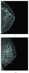
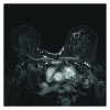
We present an interesting case of focal amyloidosis of the left breast which was intermixed with ductal carcinoma in situ (DCIS). On subsequent staging bilateral breast magnetic resonance imaging (MRI), the patient was found to have an additional suspicious enhancing mass with spiculated borders within the left breast. This mass was biopsy proven to represent pleomorphic invasive lobular carcinoma. A pulmonary nodule within the lingula was also noted on the staging bilateral breast MRI and was biopsy proven to represent extranodal Castleman's disease. Therefore, it is believed that our patient had secondary amyloidosis due to Castleman's disease.
Figures


References
-
- Rocken C, Kronsbein H, Sletten K, Roessner A, Bassler R. Amyloidosis of the breast. Virchows Archiv. 2002;440(5):527–535. - PubMed
-
- Fu K, Bassett LW. Mammographic findings of diffuse amyloidosis and carcinoma of the breast. American Journal of Roentgenology. 2001;177(4):901–902. - PubMed
-
- Sabate JM, Clotet M, Gomez A, de Las Heras P, Torrubia S, Salinas T. Radiologic evaluation of uncommon inflammatory and reactive breast disorders. Radiographics. 2005;25(2):411–424. - PubMed
-
- Sabate JM, Clotet M, Torrubia S, et al. Localized amyloidosis of the breast associated with invasive lobular carcinoma. British Journal of Radiology. 2008;81(970):e252–e254. - PubMed
-
- Altiparmak MR, Pamuk GE, Pamuk O, Dugusoy G. Secondary amyloidosis in Castleman’s disease: review of the literature and report of a case. Annals of Hematology. 2002;81(6):336–339. - PubMed
LinkOut - more resources
Full Text Sources
Other Literature Sources

