

Effect of multi-walled carbon nanotube surface modification on bioactivity in the C57BL/6 mouse model
- PMID: 23432020
- PMCID: PMC4669410
- DOI: 10.3109/17435390.2013.779757
Effect of multi-walled carbon nanotube surface modification on bioactivity in the C57BL/6 mouse model
Abstract
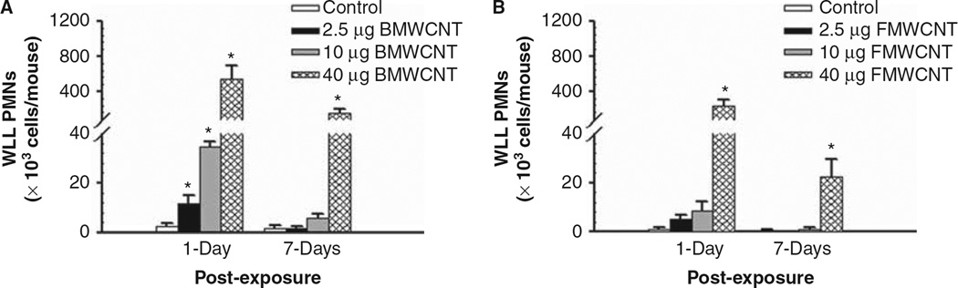
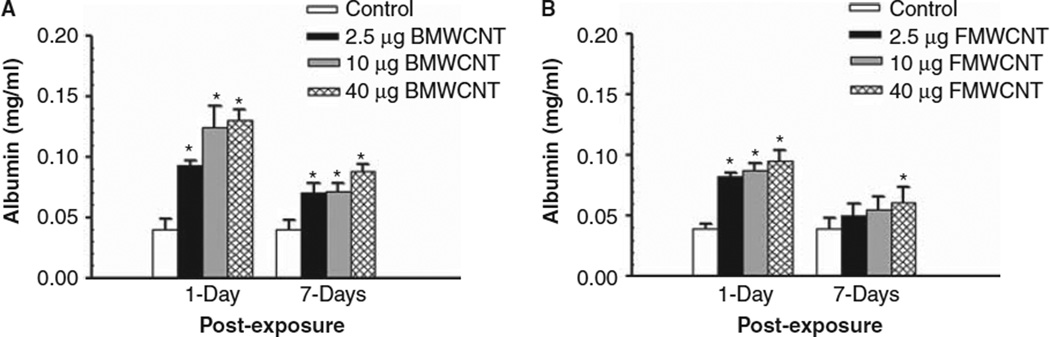
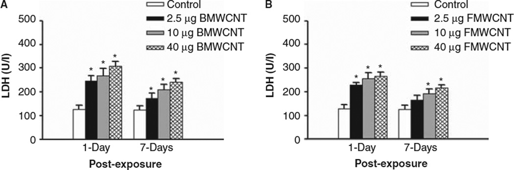
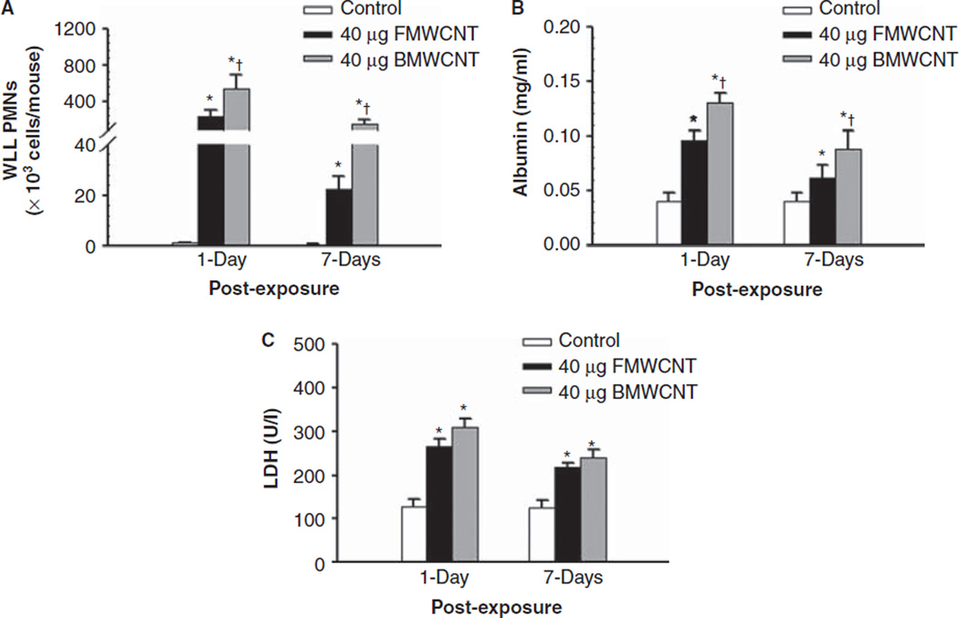
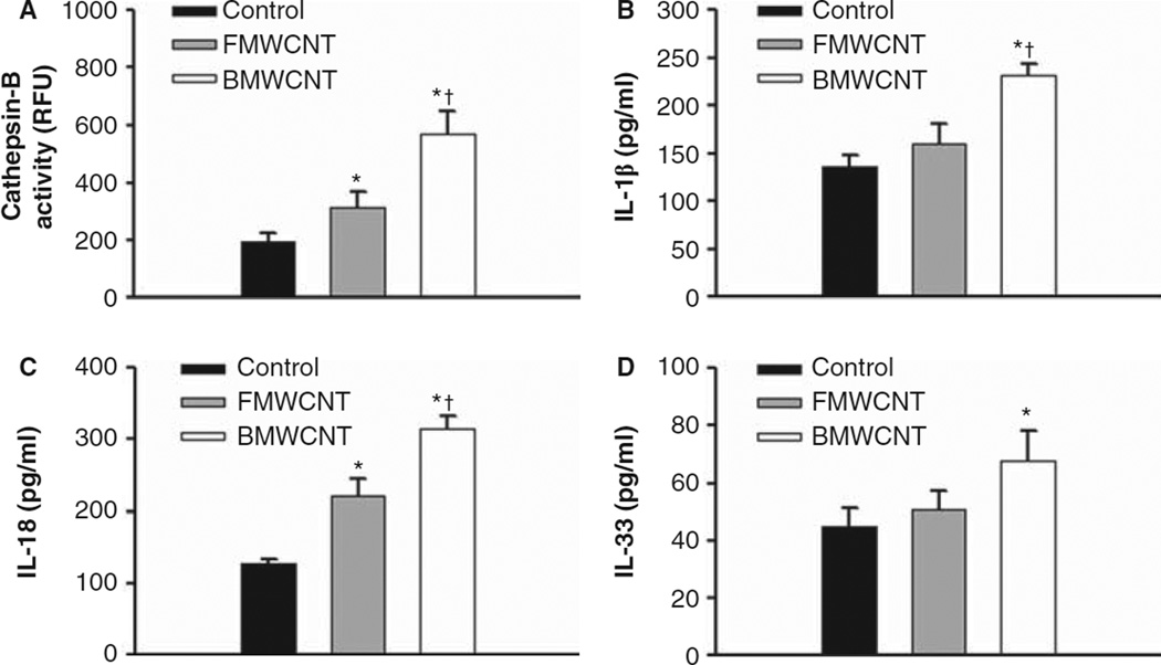
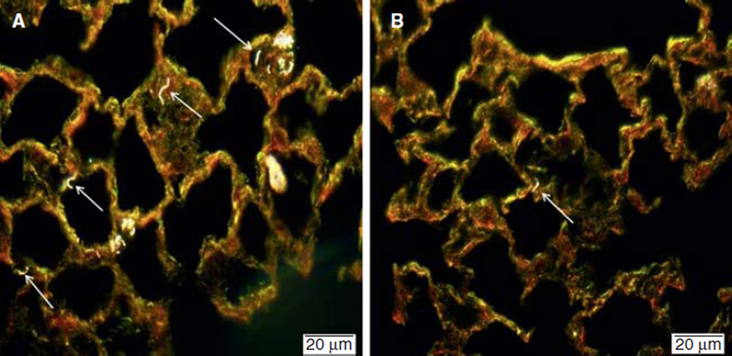
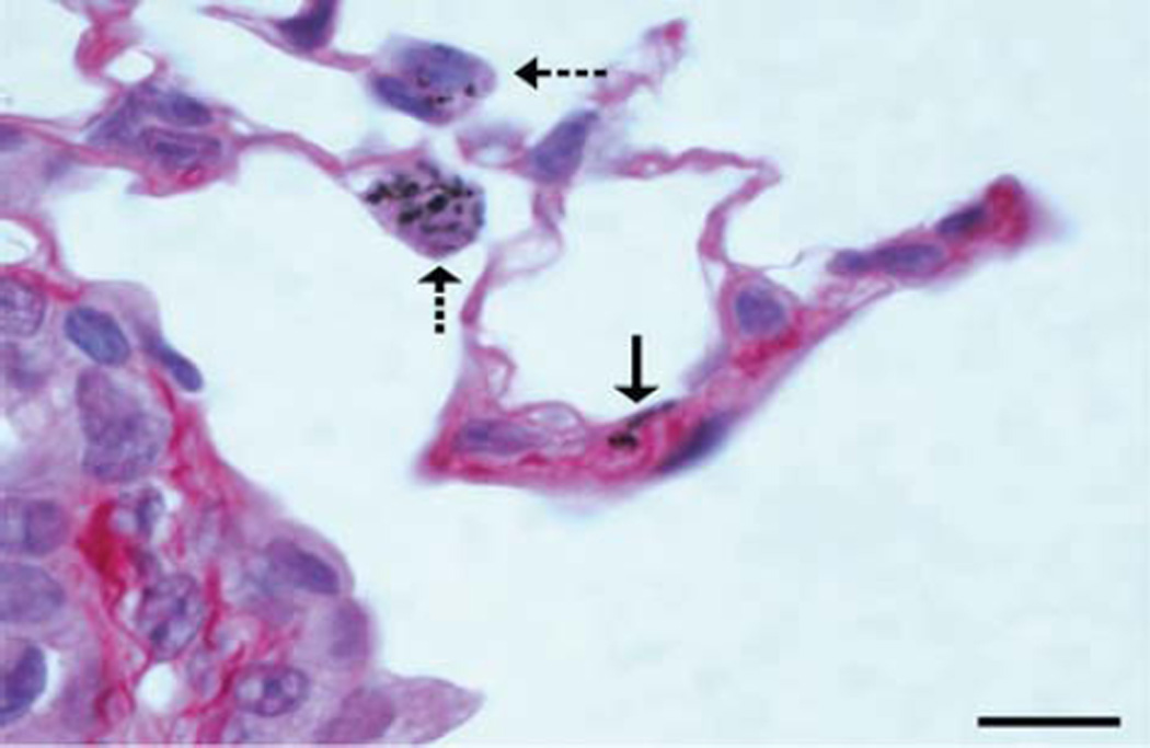
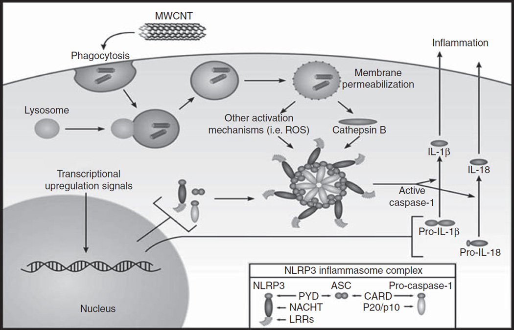
The current study tests the hypothesis that multi-walled carbon nanotubes (MWCNT) with different surface chemistries exhibit different bioactivity profiles in vivo. In addition, the study examined the potential contribution of the NLRP3 inflammasome in MWCNT-induced lung pathology. Unmodified (BMWCNT) and MWCNT that were surface functionalised with -COOH (FMWCNT), were instilled into C57BL/6 mice. The mice were then examined for biomarkers of inflammation and injury, as well as examined histologically for development of pulmonary disease as a function of dose and time. Biomarkers for pulmonary inflammation included cytokines, mediators and the presence of inflammatory cells (IL-1β, IL-18, IL-33, cathepsin B and neutrophils) and markers of injury (albumin and lactate dehydrogenase). The results show that surface modification by the addition of the -COOH group to the MWCNT, significantly reduced the bioactivity and pathogenicity. The results of this study also suggest that in vivo pathogenicity of the BMWCNT and FMWCNT correlates with activation of the NLRP3 inflammasome in the lung.
Figures
Similar articles
-
Mouse pulmonary dose- and time course-responses induced by exposure to nitrogen-doped multi-walled carbon nanotubes.Inhal Toxicol. 2020 Jan;32(1):24-38. doi: 10.1080/08958378.2020.1723746. Epub 2020 Feb 7. Inhal Toxicol. 2020. PMID: 32028803 Free PMC article.
-
Multi-walled carbon nanotube physicochemical properties predict pulmonary inflammation and genotoxicity.Nanotoxicology. 2016 Nov;10(9):1263-75. doi: 10.1080/17435390.2016.1202351. Epub 2016 Jul 7. Nanotoxicology. 2016. PMID: 27323647 Free PMC article.
-
Acute pulmonary dose-responses to inhaled multi-walled carbon nanotubes.Nanotoxicology. 2013 Nov;7(7):1179-94. doi: 10.3109/17435390.2012.719649. Epub 2012 Sep 13. Nanotoxicology. 2013. PMID: 22881873 Free PMC article.
-
NLRP3 inflammasome activation in murine alveolar macrophages and related lung pathology is associated with MWCNT nickel contamination.Inhal Toxicol. 2012 Dec;24(14):995-1008. doi: 10.3109/08958378.2012.745633. Inhal Toxicol. 2012. PMID: 23216160 Free PMC article.
-
Extracellular HMGB1 regulates multi-walled carbon nanotube-induced inflammation in vivo.Nanotoxicology. 2015 May;9(3):365-72. doi: 10.3109/17435390.2014.933904. Epub 2014 Jul 1. Nanotoxicology. 2015. PMID: 24983895 Free PMC article.
Cited by
-
Transcriptome Profile Alterations with Carbon Nanotubes, Quantum Dots, and Silver Nanoparticles: A Review.Genes (Basel). 2021 May 23;12(6):794. doi: 10.3390/genes12060794. Genes (Basel). 2021. PMID: 34070957 Free PMC article. Review.
-
Adsorption of surfactant protein D from human respiratory secretions by carbon nanotubes and polystyrene nanoparticles depends on nanomaterial surface modification and size.Philos Trans R Soc Lond B Biol Sci. 2015 Feb 5;370(1661):20140038. doi: 10.1098/rstb.2014.0038. Philos Trans R Soc Lond B Biol Sci. 2015. PMID: 25533095 Free PMC article.
-
Inhalation Exposure to Carbon Nanotubes (CNT) and Carbon Nanofibers (CNF): Methodology and Dosimetry.J Toxicol Environ Health B Crit Rev. 2015;18(3-4):121-212. doi: 10.1080/10937404.2015.1051611. J Toxicol Environ Health B Crit Rev. 2015. PMID: 26361791 Free PMC article.
-
Instillation versus inhalation of multiwalled carbon nanotubes: exposure-related health effects, clearance, and the role of particle characteristics.ACS Nano. 2014 Sep 23;8(9):8911-31. doi: 10.1021/nn503887r. Epub 2014 Aug 21. ACS Nano. 2014. PMID: 25144856 Free PMC article.
-
Effect of surface functionalizations of multi-walled carbon nanotubes on neoplastic transformation potential in primary human lung epithelial cells.Nanotoxicology. 2017 Jun;11(5):613-624. doi: 10.1080/17435390.2017.1332253. Epub 2017 Jun 2. Nanotoxicology. 2017. PMID: 28513319 Free PMC article.
References
-
- Abuilaiwi F, Laoui T, Al-Harthi M, Atieh MA. Modification and functionalization of multiwalled carbon nanotube (MWCNT) via Fischer esterification. Arabian J Sci Eng. 2010;35 Number 1C.
-
- Arend W, Palmer G, Gagay C. IL-1, IL-18, and IL-33 families of cytokines. Immunol Rev. 2008;223:20–38. - PubMed
-
- Brozena AH, Moskowitz J, Shao B, Deng S, Liao H, Gaskell KJ, et al. Outer wall selectively oxidized, water-soluble double-walled carbon nanotubes. J Am Chem Soc. 2010;132:3932–3938. - PubMed
Publication types
MeSH terms
Substances
Grants and funding
LinkOut - more resources
Full Text Sources
Other Literature Sources
Medical
Miscellaneous