. 2013 Feb 22:14:119.
doi: 10.1186/1471-2164-14-119.

MITEs in the promoters of effector genes allow prediction of novel virulence genes in Fusarium oxysporum

Affiliations

MITEs in the promoters of effector genes allow prediction of novel virulence genes in Fusarium oxysporum

Sarah M Schmidt et al. BMC Genomics. .

Abstract

Background: The plant-pathogenic fungus Fusarium oxysporum f.sp.lycopersici (Fol) has accessory, lineage-specific (LS) chromosomes that can be transferred horizontally between strains. A single LS chromosome in the Fol4287 reference strain harbors all known Fol effector genes. Transfer of this pathogenicity chromosome confers virulence to a previously non-pathogenic recipient strain. We hypothesize that expression and evolution of effector genes is influenced by their genomic context.

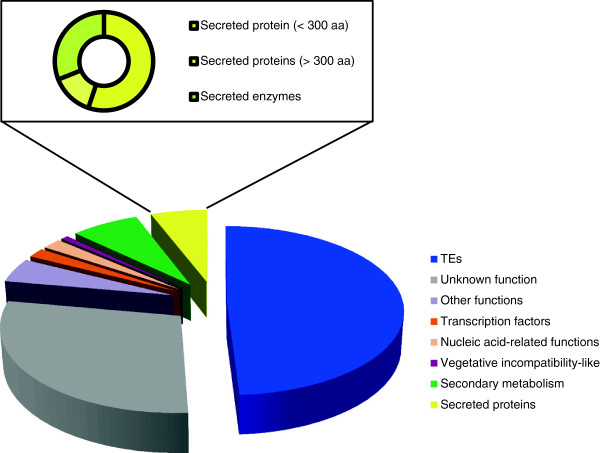
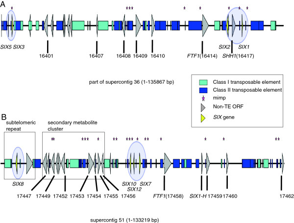
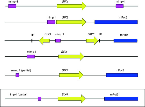
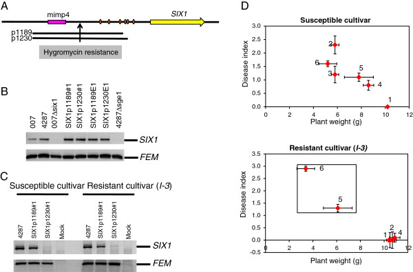
Results: To gain a better understanding of the genomic context of the effector genes, we manually curated the annotated genes on the pathogenicity chromosome and identified and classified transposable elements. Both retro- and DNA transposons are present with no particular overrepresented class. Retrotransposons appear evenly distributed over the chromosome, while DNA transposons tend to concentrate in large chromosomal subregions. In general, genes on the pathogenicity chromosome are dispersed within the repeat landscape. Effector genes are present within subregions enriched for DNA transposons. A miniature Impala (mimp) is always present in their promoters. Although promoter deletion studies of two effector gene loci did not reveal a direct function of the mimp for gene expression, we were able to use proximity to a mimp as a criterion to identify new effector gene candidates. Through xylem sap proteomics we confirmed that several of these candidates encode proteins secreted during plant infection.

Conclusions: Effector genes in Fol reside in characteristic subregions on a pathogenicity chromosome. Their genomic context allowed us to develop a method for the successful identification of novel effector genes. Since our approach is not based on effector gene similarity, but on unique genomic features, it can easily be extended to identify effector genes in Fo strains with different host specificities.


Figures

Figure 1
Figure 1
TEs dominate on the pathogenicity chromosome. TEs and non-TE genes are presented as percentage of the total TE/gene content. Genes coding for secreted proteins (including SIX genes) constitute one of the best-represented classes.
Figure 2
Figure 2
SIX genes reside in class II TE-enriched chromosomal subregions. TE densities and SIX gene locations were displayed in the IGV Genome Browser. Supercontigs are ordered according to their position in the optical map of chromosome 14, ignoring gaps between them. The positions of the SIX genes are indicated by stars. Numbers above the enlarged windows refer to position (kb) in the respective supercontig (sc).
Figure 3
Figure 3
Details of two class II TE-enriched chromosomal subregions with SIX gene mini-clusters. Schematic representations of two equally large regions of the Fol4287 pathogenicity chromosome: (A) part of supercontig 2.36 and (B) supercontig 2.51. Numbers represent FOXG gene numbers. Italic descriptions highlight interesting genes as reference points. Boxes indicate the telomeric repeat region and the putative secondary metabolite gene cluster. The genomic maps were drawn to scale.
Figure 4
Figure 4
SIX genes harbor a mimp in their promoter.SIX gene loci including 1500 bp up-and downstream of the SIX ORF are drawn to scale. A miniature Impala (mimp, pink box) is present in the promoters of SIX1 – SIX3 and SIX5 – SIX7 in Fol4287 (race 2 isolate). The SIX4 locus (boxed) is not present in the race 2 isolate. It was sequenced and analyzed in the race 1 isolate Fol004. Downstream of some SIX genes is another MITE, mFot5 (dark blue box). Inverted repeats (IR) flanking the SIX3/SIX5 locus are represented by thick black lines.
Figure 5
Figure 5
Deletion of the mimp in the SIX1 promoter does not impair SIX1 expression, but a small region with a conserved motif is required for SIX1 expression during plant infection. (A) Schematic representation of the SIX1 locus. Black lines: deleted promoter fragments (deletion length in bp); pink box: mimp; yellow arrow: SIX gene; orange circles: sequence matching AAGTCGGCAGTT[AG] motif enriched in SIX1-7 promoters. (B) In vitro expression of SIX1 in the promoter deletion strains. Mycelium of the indicated Fol strains was collected after growth in minimal medium, From the collected mycelium RNA was extracted and RT-PCR was performed to detect transcripts of SIX1 and, as control, the constitutively expressed FEM gene. (C) In planta expression of SIX1 in the promoter deletion strains. Ten days old susceptible (without resistance genes) or resistant (encoding the I-3 resistance protein that recognizes Six1) tomato seedlings were inoculated with the indicated Fol strains or with water (mock). Roots were harvested 9 dpi (days post inoculation, RNA was extracted and RT-PCR was performed as described above. (D) Disease assay of tomato plants. Ten days old seedlings of susceptible or resistant tomato seedlings (as above) were inoculated with the indicated strains or with water. Three weeks after inoculation disease was scored by determining the plant weight above the cotyledons and by phenotypic scoring according to a disease index ranging from zero (no disease) to four (heavily diseased or dead plants). (1) mock, (2) WT, (3) SIX1p1189#1, (4) SIX1p1189#2, (5) SIX1p1230#1, (6) SIX1p1230#2. Please note that, SIX1p1189#1 was not included in the bioassay with the resistant cultivar, because it is not pathogenic (see D). A black box marks interactions where recognition of Six1 by I-3 is broken or where no disease is caused. Error bars indicate the 95% confidence interval of the mean.
Figure 6
Figure 6
Deletion of the mimp in the shared promoter region of SIX3 and SIX5 does not affect expression of the two genes. (A) Schematic representation of the SIX3/SIX5 locus. Black lines: deleted promoter fragments (length of the deletion in bp; pink box: mimp; yellow arrow: SIX gene; orange circles: sequence matching AAGTCGGCAGTT[AG] motif, which is enriched in SIX1-7 promoters. (B) In vitro expression of SIX3 and SIX5 in the promoter deletion strains. Mycelium of the indicated Fol strains was collected after growth in minimal medium, RNA was extracted and RT-PCR was performed. (C) In planta expression of SIX3 and SIX5 in the promoter deletion strains. Roots of ten days old susceptible (without resistance genes) or resistant (encoding the I-2 resistance protein that recognizes Six3) tomato seedlings were inoculated with the indicated Fol strains or with water (mock). Roots were harvested 9 dpi (days post inoculation), RNA was extracted and RT-PCR was performed as described above. Expression of SIX1 was used as a control. (D) Disease assay of tomato plants. Performed as above. Error bars indicate the 95% confidence interval of the mean. Numbers indicate the strains with which the tomato plants were inoculated: (1) mock, (2) Fol007 (WT), (3) SIX3p539#2, (4) SIX3p807#1, (5) SIX3p807#2, (6) SIX3p859#1, (7) SIX3p859#2. A black box marks interactions where recognition of Six3 by I-2 is broken or where no disease is caused.

References

    1. Ma L-J, van der Does HC, Borkovich KA, Coleman JJ, Daboussi M-J, Di Pietro A, Dufresne M, Freitag M, Grabherr M, Henrissat B. Comparative genomics reveals mobile pathogenicity chromosomes in Fusarium. Nature. 2010;464(7287):367–373. doi: 10.1038/nature08850. - DOI - PMC - PubMed
    1. Rep M, Kistler HC. The genomic organization of plant pathogenicity in Fusarium species. Curr Opin Plant Biol. 2010;13(4):420–426. doi: 10.1016/j.pbi.2010.04.004. - DOI - PubMed
    1. Recorbet G, Steinberg C, Olivain C, Edel V, Trouvelot S, Dumas-Gaudot E, Gianinazzi S, Alabouvette C. Wanted: pathogenesis-related marker molecules for Fusarium oxysporum. New Phytol. 2003;159(1):73–92. doi: 10.1046/j.1469-8137.2003.00795.x. - DOI - PubMed
    1. Lievens B, Rep M, Thomma BP. Recent developments in the molecular discrimination of formae speciales of Fusarium oxysporum. Pest Manag Sci. 2008;64(8):781–788. doi: 10.1002/ps.1564. - DOI - PubMed
    1. O’Donnell K, Cigelnik E, Casper HH. Molecular phylogenetic, morphological, and mycotoxin data support reidentification of the Quorn mycoprotein fungus as Fusarium venenatum. Fungal Genet Biol. 1998;23(1):57–67. doi: 10.1006/fgbi.1997.1018. - DOI - PubMed

Publication types

MeSH terms

Substances

LinkOut - more resources