. 2013 Feb 21:10:21.
doi: 10.1186/1742-4690-10-21.

DNA damage enhances integration of HIV-1 into macrophages by overcoming integrase inhibition

Affiliations

DNA damage enhances integration of HIV-1 into macrophages by overcoming integrase inhibition

Takayoshi Koyama et al. Retrovirology. .

Abstract

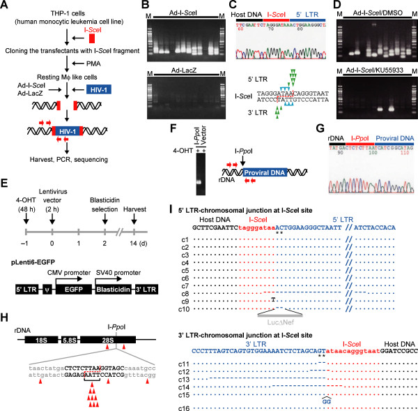
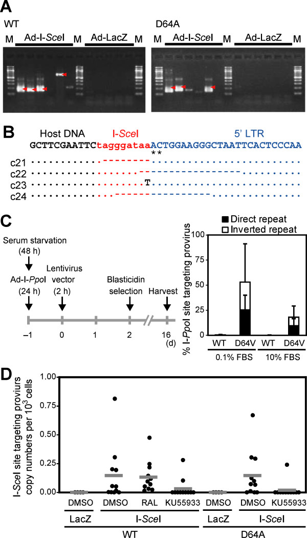
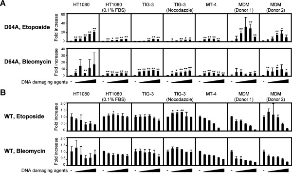
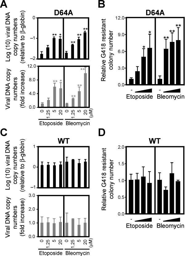
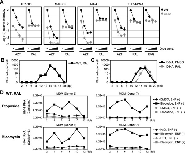
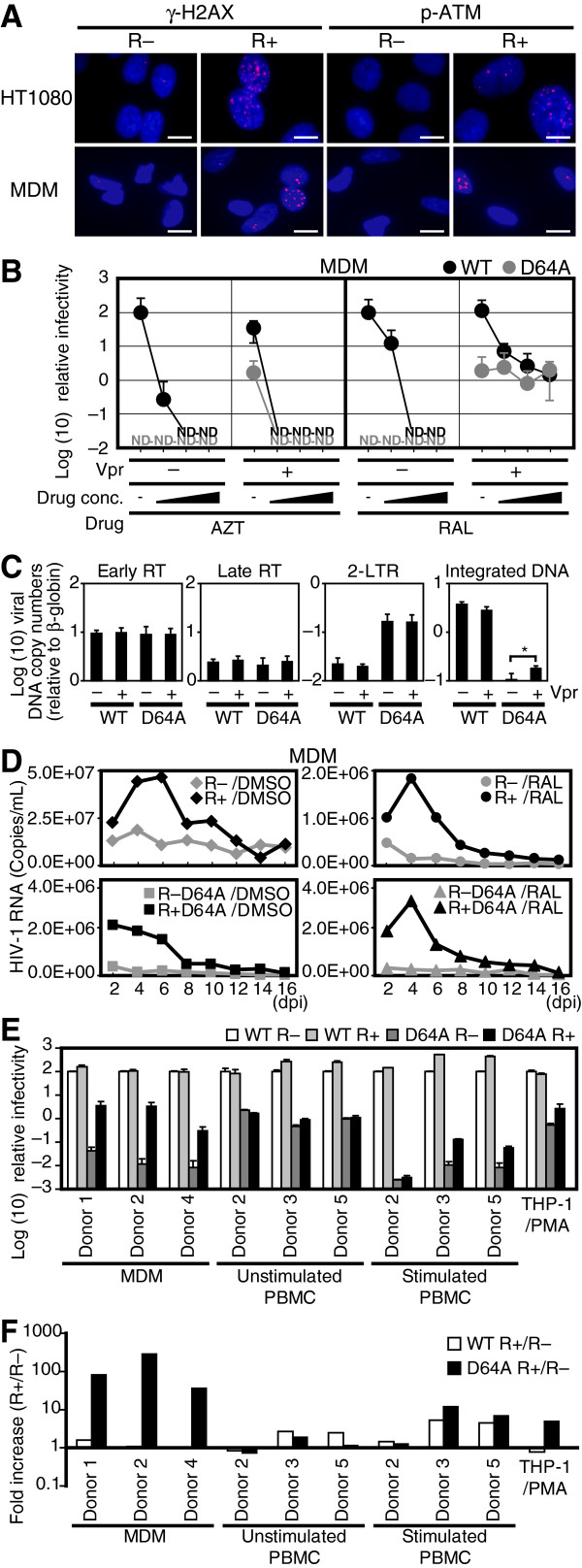
Background: The prevention of persistent human immunodeficiency virus type 1 (HIV-1) infection requires the clarification of the mode of viral transduction into resting macrophages. Recently, DNA double-strand breaks (DSBs) were shown to enhance infection by D64A virus, which has a defective integrase catalytic activity (IN-CA). However, the mechanism by which DSBs upregulate viral transduction was unclear. Here we analyzed the roles of DSBs during IN-CA-independent viral transduction into macrophages.

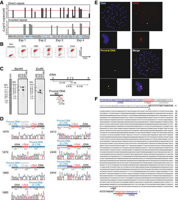
Results: We used cellular systems with rare-cutting endonucleases and found that D64A virus integrated efficiently into the sites of artificially induced DSBs. This IN-CA-independent viral transduction was blocked by an inhibitor of ataxia telangiectasia mutated protein (ATM) but was resistant to raltegravir (RAL), an inhibitor of integrase activity during strand transfer. Moreover, Vpr, an accessory gene product of HIV-1, induced DSBs in resting macrophages and significantly enhanced the rate of IN-CA-independent viral transduction into macrophages with concomitant production of secondary viruses.

Conclusion: DSBs contribute to the IN-CA-independent viral infection of macrophages, which is resistant to RAL. Thus, the ATM-dependent cellular pathway and Vpr-induced DNA damage are novel targets for preventing persistent HIV-1 infection.


Figures

Figure 1
Figure 1
HIV-1 DNA integrates into a DSB site. (A) An experimental procedures for (B)–(D) and (I). Red arrows indicate the primers used in (B) and (D). (B) PCR amplification of WT provirus DNA integrated in the I-SceI site. Each lane depicts each result of twelve samples independently infected with WT virus and Ad-I-SceI (upper panel) or Ad-LacZ (lower panel). M, molecular marker. (C) Upper panel, representative sequencing chromatogram of the PCR amplicon in samples, which were shown in upper panel of (B). Lower panel, summary of viral DNA integration sites. The 18-bp recognition sequence of the I-SceI site is shown. When digested with I-SceI, a 3-protruding end of 4 nucleotides is formed (dotted red line). Each arrowhead indicates an actual integration site of viral DNA in samples shown in (B). Integration sites were identified on most of clones except for two clones, which are indicated by arrowheads with a horizontal bar. (D) Effect of KU55933 on viral DNA integration into the I-SceI site. (E) Schematic outline of the I-PpoI-PCR experimental design in (F)–(H) (Top panel). The lentiviral vector was used in this study (bottom panel). (F) PCR amplification of lentiviral vector inserted in the I-PpoI site. Primers are shown by red arrows (G) A representative result of sequence analysis of proviral DNA integrated in the I-PpoI site. (H) Summary of integration sites of the lentiviral vector. Each arrowhead depicts each result of independent clones. The dotted line indicates I-PpoI site with a 3-protruding end of 4 nucleotides. (I) Summary of the I-SceI-PCR sequence data. A representative nucleotide sequence was shown at the top of each panel. Asterisks indicate the pAC that would be normally removed during IN-mediated integration (see Additional file 1: Figure S2). Dots indicate identical sequence to that of the representative sequence. Dashes indicate deleted nucleotides.
Figure 2
Figure 2
Frequent integration of the IN-CA defective virus into the DSB site. (A) PCR amplification of provirus DNA integrated into the I-SceI site after infection of WT virus (left) or NL-Luc-IN-D64A-E(−)R(−) virus (D64A virus) (right). PMA-treated THP-1/I-SceI cells were used. Each lane shows an independent result that was obtained from cells cultured in a single well of 6-multiwell. For each test group, six wells were independently infected with viruses. M, molecular marker. Arrowheads indicate amplicons of viral DNA integrated in the I-SceI site, which was further confirmed by sequence analysis. (B) Sequence data of D64A provirus DNA that was integrated in the I-SceI site. A representative result is shown at the top. Asterisks indicate the pAC. Dots indicate identical nucleotides to those of the representative sequence. Dashes indicate deleted nucleotides. (C) Experimental protocol for evaluating the frequency of viral integration into the DSB site. I-PpoI-qPCR and EGFP-qPCR analyses were done for quantification of I-PpoI site-specific and total proviral DNA copy numbers, respectively. Representative data of two independent experiments was shown. Error bars, s.d. of triplicate assays. (D) Evaluation of I-SceI site-targeting efficiency. PMA-treated THP-1/I-SceI cells were infected with WT or D64A virus for 2 h, and cells were harvested 48 hpi for the I-SceI-qPCR analysis (see Methods section). To cleave the I-SceI site, cells were infected with the Ad-I-SceI at an MOI of 100 from 1 h post HIV-1 infection. Treatment with RAL and KU55933 was conducted from −2 h to 48 hpi. Effects of RAL and KU55933 were evaluated on 11 samples that were prepared from three independent experiments. Each dot indicates copy numbers of provirus DNA that had integrated in the I-SceI site in 103 cells, which were infected as a single test sample.
Figure 3
Figure 3
DNA damage enhances the IN-CA independent infectivity of HIV-1. Cells were infected with D64A (A) or WT (B) viruses in the presence of etoposide or bleomycin from 0–48 h post-infection (hpi). After 48 h, cell extracts were prepared and subjected to the luciferase assay. The fold increase of the activity after each viral infection with or without DNA damaging agents was shown. In experiments using cell lines, representative data from one of repeated experiments was shown. Results are presented as mean ± s.d. of triplicate assays. All cells except for MT-4 cells were treated with 0, 0.625, 1.25, 2.5, 5, 10 μM etoposide or 0, 1.25, 2.5, 5, 10, 20 μM bleomycin. MT-4 cells were treated with 0, 0.039, 0.078, 0.156, 0.313, 0.625 μM etoposide or 0, 0.078, 0.156, 0.313, 0.625, 1.25 μM bleomycin. Raw data of luciferase activity was shown in Additional file 1: Figure S4. *, P < 0.05; **, P < 0.01.
Figure 4
Figure 4
DNA damage enhances the integration rate of HIV-1. Serum-starved HT1080 cells were infected with D64A (A) or WT (C) viruses in the presence of etoposide or bleomycin from 0–24 hpi. After 48 h, genomic DNA was extracted and subjected to qPCR. Relative copy numbers of HIV-1 DNA to β-globin were estimated (top) and the fold increase of HIV-1 DNA copy number compared to control infection that was conducted without DNA damaging agents (bottom) were calculated. For colony formation assay, VSVG-pseudotyped D64A (NL-Neo-IN-D64A-E(−)R(−)) (B) or WT (NL-Neo-E(−)R(−)) (D) viruses, which had the neomycin resistant gene (NeoR), were used. HT1080 cells were treated with various doses of etoposide or bleomycin for 24 h, which were added at the same time of viral infection. After selection with 600 μg/mL of G418, numbers of NeoR colonies were counted. Numbers of NeoR colonies were normalized by plating efficiency. Error bars, s.d. of triplicate assays. *P < 0.05; **P < 0.01.
Figure 5
Figure 5
IN-CA deficient virus can produce infectious progeny virus. (A) Effect of RAL on the infectivity of WT and D64A viruses. After infection with VSVG-pseudotyped WT (NL-Luc-E(−)) or D64A (NL-Luc-IN-D64A-E(−)) virus in the presence of AZT, RAL or EVG, cells were harvested at 48 hpi and subjected to luciferase assay. Relative luciferase activity compared to a control sample, in which WT virus was infected without any compounds, were plotted. Concentration of AZT was 0, 1, 10 and 100 μM, whereas concentrations of RAL and EVG were 0, 0.1, 1 and 10 μM. Black circles, WT virus; gray circles, D64A virus; ND, not detected. Representative data of two independent experiments was shown. Error bars, s.d. of triplicate assays. (B) Functional evaluation of progeny viruses generated after IN-CA independent infection. MT-4 cells were infected in the presence of RAL with replication-competent WT virus (NL4-3). Then conditioned medium was harvested every 2 d, and infectivity of progeny virus present in the conditioned medium was evaluated using MAGIC5 cells. (C) Similar experiment with (B) was done using D64A (NL-IN-D64A) virus. Representative results of three independent experiments are shown. (D) Secondary virus generated from MDMs infected with virus in the presence of RAL. MDMs were infected with a replication-competent NL4-3 virus with an env gene, which was derived from R5-tropic ADA virus (NL-ADA-R(−)). Then, HIV-1 RNA copy number in the conditioned medium was quantified by RT-qPCR analysis. To evaluate effects of DNA damaging agents, 2.5 μM etoposide or 1.25 μM bleomycin were added from 0–2 dpi. To exclude the possibility that carry-over virions, which were remnant viruses that could not be completely removed after the initial infection, we included control sample, in which a fusion inhibitor enfuvirtide (ENF) was continuously added from 0 dpi to the end of assay.
Figure 6
Figure 6
Detection of intact provirus DNA in the DSB site. (A) I-PpoI-qPCR screening of cell clones containing provirus DNA in I-PpoI site. HT1080 cells were infected with Ad-I-PpoI at an MOI of 30 in the medium with 0.1% FBS. After 24 h, cells were further infected with lentiviruses (VSVG-pseudotyped Lenti6-EGFP-D64V) also under serum-starved conditions. Two h later, medium was changed with fresh one with 0.1% FBS. On the next day, medium was replaced with a complete medium with 10% FBS. Blasticidin-resistant colonies were isolated and I-PpoI site targeting provirus was detected by I-PpoI-qPCR. The threshold of detecting provirus integrated as direct or inverted repeat orientation was −1 log(10) copies/cell (indicated in red horizontal lines). (B) EGFP expression analysis. Cells containing the proviral DNA in I-PpoI site in (A) were further analyzed for the expression of EGFP by flow cytometer. (C) The estimation of proviral DNA copy number. Copy numbers of provirus DNA in EGFP-positive clones, shown in (B), were analyzed by Southern blot by using a part of DNA fragment of the lentiviral vector as a probe. Genomic DNA extracted from each clone was digested with BamHI or EcoRI prior to electrophoresis. Restriction maps are shown (right panel). B, BamHI; E, EcoRI; P, I-PpoI. Of note, clone #2413 possessed a single copy of provirus DNA. (D) Sequence analysis of lentiviral vector integrated in the I-PpoI site. EGFP-positive clones shown in (B) were subjected to sequence analysis. I-PpoI-PCR amplicons were directly used as a template for sequence analysis. (E) FISH analysis of the #2413 clone. (F) Nucleotide sequence of intact proviral DNA present in the DSB site. The proviral DNA of #2413 clone was sequenced and whole nucleotide sequence data was shown. In #2413 clone, no structural alternations of provirus DNA were detected.
Figure 7
Figure 7
Vpr mimics DNA damaging agents, and enhances the IN-CA independent macrophage infection. (A) Vpr induces DNA damage cellular signals in MDMs. HT1080 cells or MDMs were infected with VSVG-pseudotyped R– virus (NL-Luc-E(−)R(−)) or R+ virus (NL-Luc-E(−)), and then analyzed immunochemically. Bars = 10 μm. (B) Effect of RAL on the infectivity of WT and D64A viruses. MDMs were infected with WT or D64A viruses in the presence of AZT or RAL. The cells were harvested at 48 hpi and subjected to luciferase assay. Relative luciferase activity values to WT R– infectivity are shown. Black circles, WT; gray circles, D64A; ND, not detected. Error bars, s.d. of triplicate assays. (C) Effects of Vpr on the integration of viral DNA into the host genome. Serum-starved HT1080 cells were infected with VSVG-pseudotyped IN WT or D64A mutant virus with or without Vpr. After 48 h, infected cells were subjected to qPCR analysis. Error bars, s.d. of triplicate assays. *P < 0.05. (D) HIV-1 replicates in MDMs in the presence of RAL. Replication-competent NL4-3 with an intact env gene derived from R5-tropic ADA viruses (NL-ADA, NL-ADA-R(−), NL-ADA-IN-D64A, and NL-ADA-IN-D64A-R(−)) were infected. Then, copy numbers of HIV-1 RNA in the conditioned medium was quantified by RT-qPCR. E and F) Positive effects of Vpr on infection of D64A virus into MDMs. Primary cells and cell lines were infected with IN WT or D64A mutant virus with or without Vpr. Cells were harvested at 48 hpi and subjected to luciferase assay. (E) Relative luciferase activity values to WT R– infectivity are shown. White bars, WT/R–; light gray bars, WT/R+; dark gray bars, D64A/R–; black bars, D64A/R+. Error bars, s.d. of triplicate assays. (F) Fold increase of R+ virus infectivity to R– virus. White bars, WT; black bars, D64A.

Similar articles

Cited by

References

    1. Perelson AS, Essunger P, Cao Y, Vesanen M, Hurley A, Saksela K, Markowitz M, Ho DD. Decay characteristics of HIV-1-infected compartments during combination therapy. Nature. 1997;387:188–191. doi: 10.1038/387188a0. - DOI - PubMed
    1. Richman DD, Margolis DM, Delaney M, Greene WC, Hazuda D, Pomerantz RJ. The challenge of finding a cure for HIV infection. Science. 2009;323:1304–1307. doi: 10.1126/science.1165706. - DOI - PubMed
    1. Hazuda DJ, Felock P, Witmer M, Wolfe A, Stillmock K, Grobler JA, Espeseth A, Gabryelski L, Schleif W, Blau C, Miller MD. Inhibitors of strand transfer that prevent integration and inhibit HIV-1 replication in cells. Science. 2000;287:646–650. doi: 10.1126/science.287.5453.646. - DOI - PubMed
    1. Dorr P, Westby M, Dobbs S, Griffin P, Irvine B, Macartney M, Mori J, Rickett G, Smith-Burchnell C, Napier C. Maraviroc (UK-427,857), a potent, orally bioavailable, and selective small-molecule inhibitor of chemokine receptor CCR5 with broad-spectrum anti-human immunodeficiency virus type 1 activity. Antimicrob Agents Chemother. 2005;49:4721–4732. doi: 10.1128/AAC.49.11.4721-4732.2005. - DOI - PMC - PubMed
    1. Jager S, Cimermancic P, Gulbahce N, Johnson JR, McGovern KE, Clarke SC, Shales M, Mercenne G, Pache L, Li K. Global landscape of HIV-human protein complexes. Nature. 2012;481:365–370. - PMC - PubMed

Publication types

MeSH terms

Substances