Review
. 2013 Jul;15(7):1070-8.
doi: 10.1111/cmi.12128. Epub 2013 Mar 14.

The 'ubiquitous' reality of vector immunology

Affiliations
Review

The 'ubiquitous' reality of vector immunology

Maiara S Severo et al. Cell Microbiol. 2013 Jul.

Abstract

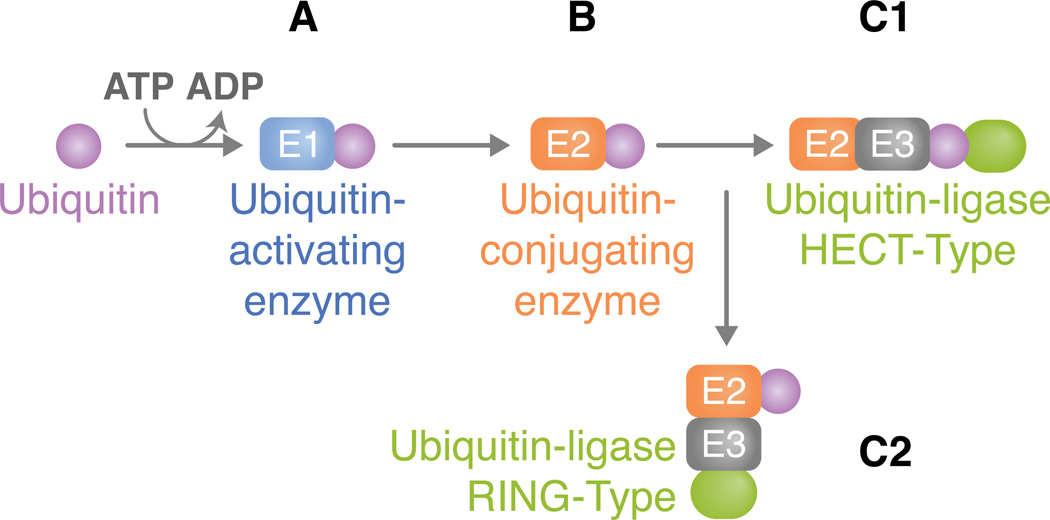
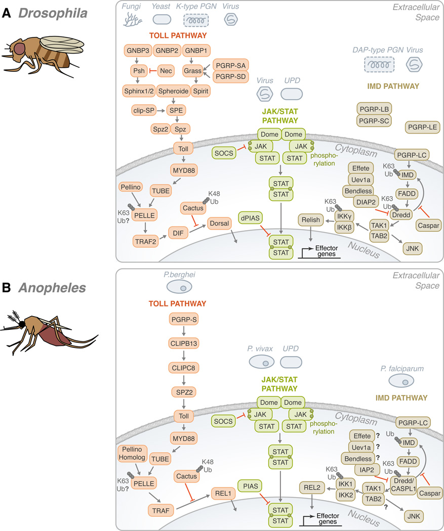
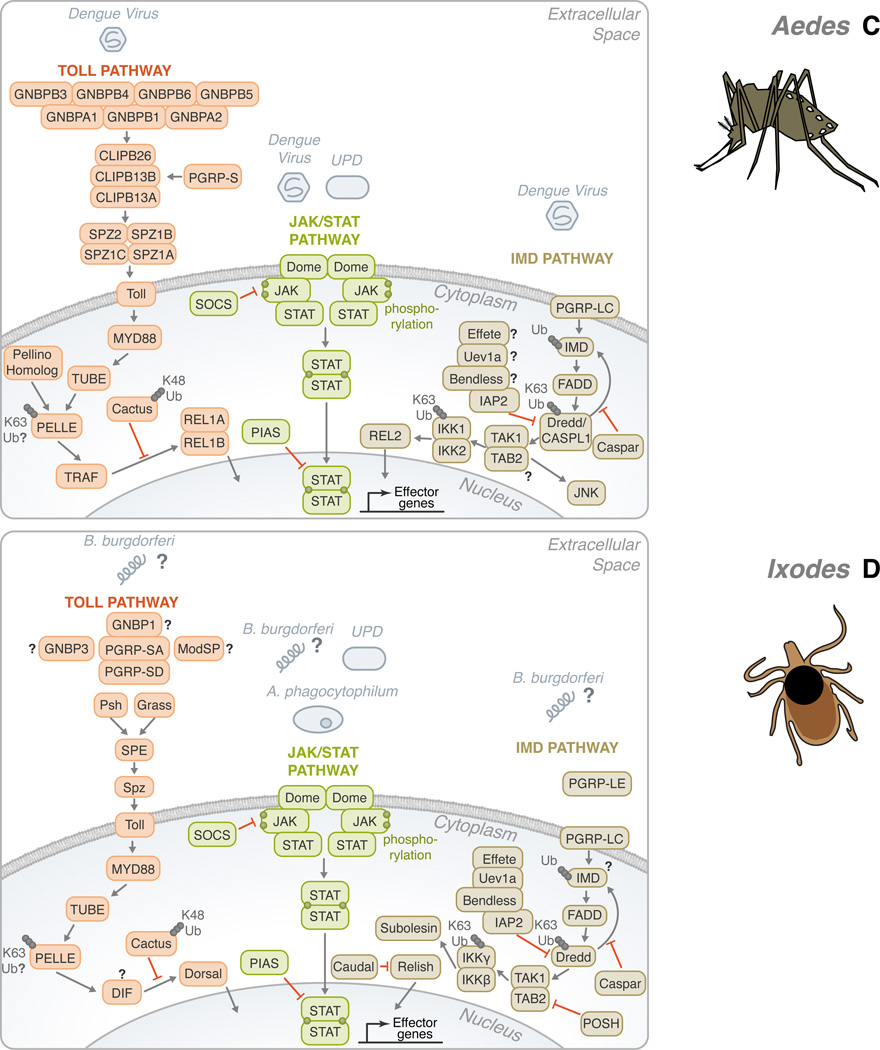
Ubiquitination (ubiquitylation) is a common protein modification that regulates a multitude of processes within the cell. This modification is typically accomplished through the covalent binding of ubiquitin to a lysine residue onto a target protein and is catalysed by the presence of three enzymes: an activating enzyme (E1), ubiquitin-conjugating enzyme (E2) and ubiquitin-protein ligase (E3). In recent years, ubiquitination has risen as a major signalling regulator of immunity and microbial pathogenesis in the mammalian system. Still, little is known about how ubiquitin relates specifically to vector immunology. Here, we provide a brief overview of ubiquitin biochemistry and describe how ubiquitination regulates immune responses in arthropods of medical relevance. We also discuss scientific gaps in the literature and suggest that, similar to mammals, ubiquitin is a major regulator of immunity in medically important arthropods.


Conflict of interest statement

The authors declare no conflict of interest.

Figures

Figure 1
Figure 1. The ubiquitination pathway
(A) For an ubiquitin to be added to a target lysine, a thiol ester bond between the E1 and ubiquitin is first formed in a two-step ATP-dependent reaction. Ubiquitin is activated through its combination with MgATP. This, in turn, forms a ubiquitin adenylate intermediate that acts as a ubiquitin donor to the E1 active site, creating an E1 molecule that consists of both an activated thiol ester ubiquitin and an adenylated ubiquitin. (B) The thiol-linked ubiquitin is then transferred to the E2. E2s typically share a core domain of about 150 amino acids and a cysteine residue in the active site to which E1 ubiquitin is transferred. (C) The ubiquitin ligase is typically a protein complex that attaches to both the substrate and the E2. The method in which substrate binding occurs varies between direct interactions or through auxiliary proteins. E3 enzymes can be classified according to the domains they carry. Here, we address two types of ubiquitin ligases: (C1) HECT type or (C2) RING type. (C1) HECT domain ligases have a domain that is approximately 350 residues. They serve a catalytic role in the conjugation of ubiquitin to the target protein (green oval). HECT domain proteins possess a conserved cysteine residue through which the activated E2 ubiquitin can form a thiol ester bond, thus leading to its transfer from E2. After attachment of ubiquitin, a lysine residue of the target protein must be introduced to the active site in order for substrate-ubiquitin ligation to occur. Ubiquitin is ultimately conjugated to a NH2 group of the target protein. The difference in the NH2 terminal domain within various HECT domain proteins is responsible for E3 substrate specificity. (C2) Unlike the HECT domain ligase that serves as a direct intermediate between E2 and substrate ubiquitin transfer, the RING type functions as a molecular scaffold that brings proteins together instead of acting as a chemical catalyst. It is defined by a conserved series of histidine and cysteine residues that is patterned to form a cross-brace structure, which permits the binding of two zinc cations.
Figure 2
Figure 2. Conservation of the Toll, IMD, and JAK/STAT pathways in Drosophila melanogaster, Anopheles gambiae, Aedes aegypti and Ixodes scapularis
(A) In Drosophila, gram-negative bacteria binding proteins (GNBPs) and peptidoglycan recognition proteins (PGRPs) have been shown to activate the Toll pathway in the presence of stimulants, such as fungi, yeast, lysine-type peptidoglycan (K-type PGN) and viruses. These recognition proteins signal downstream to Persephone (psh) and Grass. CLIP domain serine proteases (clip-SP) modulate the signaling after recognition as well. Sphinx1/2, Spheroide, and Spirit initiate the activation of Spätzle through the Spätzle processing enzyme (SPE). Spätzle binds to Toll which recruits three Death domain-containing molecules: MyD88, Tube, and Pelle. Pellino/Pellino homolog, perhaps, acts as a positive regulator of immunity by ubiquitinating Pelle. After which, TNF-receptor-associated factor (TRAF) signals to Dorsal-immune related factor (DIF), followed by signaling to Dorsal. The activation is facilitated by the degradation of Cactus through K48 ubiquitination. The transcription factor translocates to the nucleus in order to upregulate immune genes. The Toll pathway is highly conserved in: (B) Anopheles, (C) Aedes, and (D) Ixodes. The Toll pathway has been demonstrated to recognize Plasmodium berghei in (B) Anopheles and the Dengue virus in (C) Aedes. TRAF signaling initiates REL1 and REL1A /1B activity in (B) Anopheles and (C) Aedes, respectively. (A) On the other hand, the Drosophila IMD pathway recognizes primarily mono-diaminopimelic acid-type peptidoglycans (DAP-type PGN). Fas-associated protein with death domain (FADD) is recruited to IMD. FADD binds to the Death related ced-3/Nedd2-like caspase (Dredd)/CASPL1. Dredd can cleave IMD. Inhibitor of apoptosis (IAP) can also associate with Dredd/CASPL1. Effete, Uev1a, and Bendless play a role in the regulation of this step and caspar may also inhibit the activity of IMD-dependent transcription factors. TGF-β activated kinase (TAK1), TAK1 binding protein 2 (TAB2) complex forms as signaling continues. Two avenues may result from the IMD pathway: JNK or NF-κB. For NF-κB activation, Relish translocates to the nucleus to activate effector genes. There are several potential sites of ubiquitination throughout the IMD pathway: IMD, Dredd/CASPL1, and the IKK complex. Like the Toll pathway, the IMD pathway is found in many species: (B) Anopheles, (C) Aedes, and (D) Ixodes. P. falciparum and the dengue virus can trigger the IMD pathway in (B) Anopheles and (C) Aedes, respectively. While Relish is regulated by the IMD pathway in (A) Drosophila and (D) Ixodes, REL2, the homolog of Relish, acts as the transcription factor in (B) Anopheles and (C) Aedes. The third pathway is the JAK/STAT pathway. (A) A ligand derived from the unpaired (UPD) gene activates the pathway by binding to Domeless (Dome). Phosphorylated JAK promotes the dimerization of STAT. Dimerized STAT can proceed to the nucleus. Countering the activation, both SOCS and PIAS negatively regulate the JAK/STAT pathway. Though the JAK/STAT pathway is evolutionarily conserved across the organisms discussed, various pathogens have demonstrated the ability to activate the JAK-STAT pathway, such as: (B) Plasmodium vivax, (C) dengue virus and (D) A. phagocytophilum. For the Toll, IMD and JAK/STAT pathways, B. burgdorferi recognition in I. scapularis remains mostly elusive. The information illustrated here was obtained from gene sequences available at vectorbase (www.vectorbase.org) and the following articles: (Waterhouse et al., 2007, Liu et al., 2012, Souza-Neto et al., 2009, Cirimotich et al., 2010, Xi et al., 2008, Hoffmann, 2003).

References

    1. Agaisse H, Perrimon N. The roles of JAK/STAT signaling in Drosophila immune responses. Immunol Rev. 2004;198:72–82. - PubMed
    1. Almazan C, Lagunes R, Villar M, Canales M, Rosario-Cruz R, Jongejan F, de la Fuente J. Identification and characterization of Rhipicephalus (Boophilus) microplus candidate protective antigens for the control of cattle tick infestations. Parasitology Res. 2010;106:471–479. - PMC - PubMed
    1. Anderson KV, Jurgens G, Nusslein-Volhard C. Establishment of dorsal-ventral polarity in the Drosophila embryo: genetic studies on the role of the Toll gene product. Cell. 1985;42:779–789. - PubMed
    1. Arbouzova NI, Zeidler MP. JAK/STAT signalling in Drosophila: insights into conserved regulatory and cellular functions. Development. 2006;133:2605–2616. - PubMed
    1. Arjona A, Ledizet M, Anthony K, Bonafe N, Modis Y, Town T, Fikrig E. West Nile virus envelope protein inhibits dsRNA-induced innate immune responses. J Immunol. 2007;179:8403–8409. - PubMed

Publication types

MeSH terms

Substances

LinkOut - more resources