

Review
doi: 10.1016/j.ijrobp.2013.01.011.
Epub 2013 Feb 20.
Detection and repair of ionizing radiation-induced DNA double strand breaks: new developments in nonhomologous end joining
Affiliations
- PMID: 23433795
- PMCID: PMC3731128
- DOI: 10.1016/j.ijrobp.2013.01.011
Item in Clipboard
Review
Detection and repair of ionizing radiation-induced DNA double strand breaks: new developments in nonhomologous end joining
Int J Radiat Oncol Biol Phys.
.
Abstract
DNA damage can occur as a result of endogenous metabolic reactions and replication stress or from exogenous sources such as radiation therapy and chemotherapy. DNA double strand breaks are the most cytotoxic form of DNA damage, and defects in their repair can result in genome instability, a hallmark of cancer. The major pathway for the repair of ionizing radiation-induced DSBs in human cells is nonhomologous end joining. Here we review recent advances on the mechanism of nonhomologous end joining, as well as new findings on its component proteins and regulation.
Copyright © 2013 Elsevier Inc. All rights reserved.
Figures

A model for NHEJ, showing the three basic steps (end detection, processing and ligation) along with the proteins implicated in each stage. Interactions are shown by black lines with solid arrowheads. Putative interactions are in dashed lines. Blue lines indicate phosphorylation events. Blue, red and black Ps enclosed in circles indicate DNA-PK, ATM and CK2-mediated phosphorylation events, respectively. The AP lyase activity of Ku is indicated by the red arrow. For simplicity, the interaction between Artemis and DNA ligase IV [49,50] is not shown. See text for details.
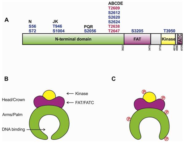
A.Schematic of DNA-PKcs showing major in vivo phosphorylation sites discussed in text. Residues in red are equivalent to residues mutated in the 3A mice [66]. Domain boundaries are shown by vertical numbers. See also [37,62,67]. B. Model for structure of DNA-PKcs, adapted from [36]. C. Model for putative effects of phosphorylation on DNA-PKcs structure, adapted from [37].
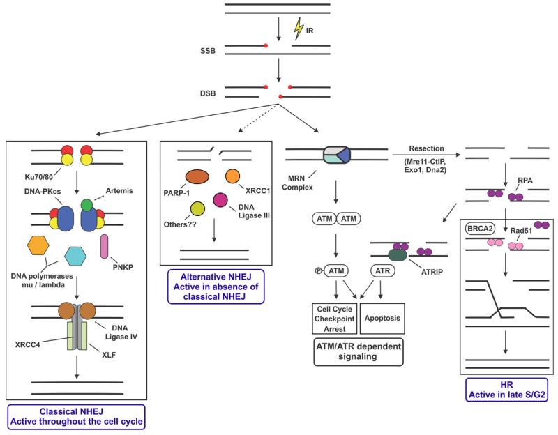
A model for the DNA damage response in mammalian cells indicating competition of DSB ends by classical NHEJ, Alt-NHEJ and HR. Adapted from [139] with permission.
References
-
- Gottlieb TM, Jackson SP. The DNA-dependent protein kinase: Requirement for DNA ends and association with ku antigen. Cell. 1993;72:131–142. - PubMed
Publication types
MeSH terms
Substances
Grants and funding
LinkOut - more resources
Full Text Sources
Other Literature Sources
