. 2013 Apr;9(4):271-6.
doi: 10.1038/nchembio.1185. Epub 2013 Feb 24.

Allosteric inhibition of hypoxia inducible factor-2 with small molecules

Affiliations

Allosteric inhibition of hypoxia inducible factor-2 with small molecules

Thomas H Scheuermann et al. Nat Chem Biol. 2013 Apr.

Abstract

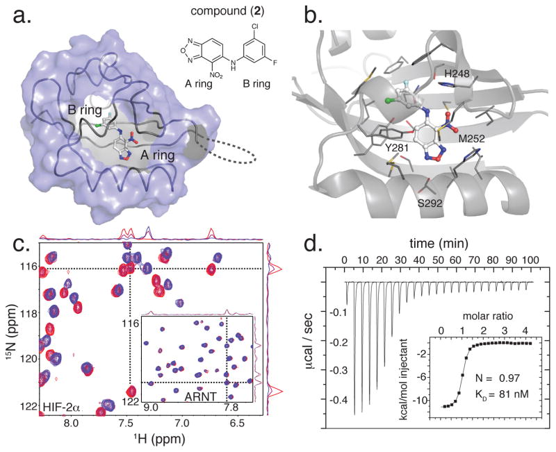
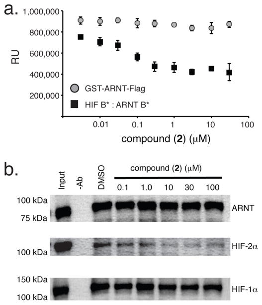
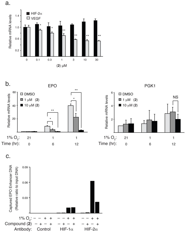
Hypoxia inducible factors (HIFs) are heterodimeric transcription factors induced in many cancers where they frequently promote the expression of protumorigenic pathways. Though transcription factors are typically considered 'undruggable', the PAS-B domain of the HIF-2α subunit contains a large cavity within its hydrophobic core that offers a unique foothold for small-molecule regulation. Here we identify artificial ligands that bind within this pocket and characterize the resulting structural and functional changes caused by binding. Notably, these ligands antagonize HIF-2 heterodimerization and DNA-binding activity in vitro and in cultured cells, reducing HIF-2 target gene expression. Despite the high sequence identity between HIF-2α and HIF-1α, these ligands are highly selective and do not affect HIF-1 function. These chemical tools establish the molecular basis for selective regulation of HIF-2, providing potential therapeutic opportunities to intervene in HIF-2-driven tumors, such as renal cell carcinomas.


Conflict of interest statement

Competing financial interests

R.K.B., K.H.G., T.H.S., J.K. and U.K.T. have received stock options and other financial compensation from Peloton Therapeutics Inc.

Figures

Figure 1
Figure 1. Biophysical characterization of the HIF-2α PAS-B-2 complex
(a) The crystal structure of the ternary complex of HIF-2 PAS-B* with compound (2) reveals ligand binding into the internal cavity sequestered from bulk solvent within the HIF-2α PAS-B domain (gray). For clarity, the ARNT-PAS-B* portion of the protein heterodimer is not shown, and a portion of the HIF-2α PAS-B* surface (blue) has been cut away to reveal the internal binding site. (b) Protein-ligand contacts as revealed by expanded view of the compound (2) binding site, showing that it is composed of a mix of polar and hydrophobic residues. (c) 15N/1H HSQC spectra of 200 μM 15N HIF-2α PAS-B (main panel) and 15N-ARNT PAS-B (inset) in the presence of 0, 125 and 250μM (2) (red to blue) demonstrate the specific binding of compound (2) to HIF-2α PAS-B. One-dimensional traces of spectra (at locations shown by dashed lines) demonstrate slow exchange binding behavior of (2) to HIF-2α, and no binding to ARNT PAS-B. (d) ITC measurements of (2) to HIF-2α PAS-B quantitate the binding affinity and 1:1 stoichiometry.
Figure 2
Figure 2. Binding of (2) into HIF-2α PAS-B affects the heterodimeric β-sheet interface between HIF PAS domains
(a) Backbone 1H and 15N chemical shift differences between apo-and (2)-bound states are mapped onto the HIF-2α PAS-B primary and secondary structures. The yellow-to-red color scale shown on the right is used in (b) and Supplementary Figure 6. (b) Ligand-induced chemical shift perturbations are mapped onto the HIF-2α PAS-B structure with spheres denoting HIF-2α Cα sites within 8 Å of ARNT PAS-B. View is approximately 180° rotated about the y (vertical) axis from the view in Figure 1a. (c) Ligand-induced conformational changes in similar regions are also evident from X-ray diffraction data, as revealed by a Fo(liganded) -Fo(apo) electron density difference map (rendered at 4σ; positive density in green, negative density in red).
Figure 3
Figure 3. Compound (2) disrupts HIF-2 heterodimerization in vitro
(a) Addition of (2) blocks heterodimer assembly between purified recombinant HIF-2α PAS-B* and ARNT PAS-B* heterodimer (squares) as assessed in the AlphaScreen Assay. No effect was observed in control reactions employing a single (doubly-tagged) GST-ARNT-PAS-B*-FLAG protein capable of recruiting both beads to induce an AlphaScreen signal (circles). Assays were performed in triplicate and the error bars represent ± SD. RU = relative units. (b) Compound (2) disrupts heterodimerization of the full length HIF-2 transcription factor. Nuclear extracts prepared from hypoxic Hep3B cells expressing ARNT, HIF-1α and HIF-2α (input) were incubated with increasing concentrations of (2). Immunoblot analysis indicates amounts of HIF polypeptides immunoprecipitated in the absence (-Ab) or presence of an anti-ARNT antibody.
Figure 4
Figure 4. Compound (2) binds selectively to HIF-2α over HIF-1α PAS-B
(a) Comparison of internal cavity sizes (blue) identified by a 1.4 Å probe within our HIF-2α PAS-B crystal structure (PDB code 3F1P) (top) and a homology model of HIF-1α PAS-B domain based on this structure. Sequence differences amongst these two closely related paralogs reduce the expected size of the HIF-1α PAS-B cavity. (b) The HIF-1α PAS-B model suggests that several sequence differences among these paralogs leads to the placement of bulkier side chains (red) within the HIF-1α PAS-B core. These substitutions appear to shrink the cavity observed in HIF-2α PAS-B (HIF-2α PAS-B cavity rendered as a blue surface, superimposed on the HIF-1α PAS-B model). Amino acid differences are indicated with the first designating HIF-2 amino acid identity, and HIF-1 identity by the last letter. (c) ITC measurements of a HIF-1α PAS-B-compound (2) titration does not show detectable protein-ligand interaction under the same conditions used to observe binding with HIF-2α PAS-B (Fig. 1d).
Figure 5
Figure 5. Compound (2) selectively antagonizes HIF-2 activity in cultured cells
While incubation of (2) with normoxic 786-0 cells has no effect on HIF-2α expression (a), RT-PCR reveals that expression of HIF-2 target genes are antagonized by (2) in both 786-0 (a) and Hep3B (b) cells. (c) Compound (2) selectively disrupts DNA binding by HIF-2, but not HIF-1, in a ChIP assay. The RT-PCR data for each gene are the mean of three values determined from three independently harvested sets and the error bars represent ± SD. Differences between paired values are statistically significant as determined by t-test. * = p < 0.01; ** = p < 0.001

References

    1. Semenza GL. Hypoxia-inducible factors: mediators of cancer progression and targets for cancer therapy. Trends Pharmacol Sci. 2012;33:207–214. - PMC - PubMed
    1. Xia X, et al. Integrative analysis of HIF binding and transactivation reveals its role in maintaining histone methylation homeostasis. Proc Natl Acad Sci U S A. 2009;106:4260–4265. - PMC - PubMed
    1. Schodel J, et al. High-resolution genome-wide mapping of HIF-binding sites by ChIP-seq. Blood. 2011;117:e207–217. - PMC - PubMed
    1. Majmundar AJ, Wong WJ, Simon MC. Hypoxia-inducible factors and the response to hypoxic stress. Mol Cell. 2010;40:294–309. - PMC - PubMed
    1. Greer SN, Metcalf JL, Wang Y, Ohh M. The updated biology of hypoxia-inducible factor. EMBO J. 2012;31:2448–2460. - PMC - PubMed

Publication types

MeSH terms

Substances