. 2013 Apr;14(4):364-71.
doi: 10.1038/ni.2541. Epub 2013 Feb 24.

The transcription factor STAT5 is critical in dendritic cells for the development of TH2 but not TH1 responses

Affiliations

The transcription factor STAT5 is critical in dendritic cells for the development of TH2 but not TH1 responses

Bryan D Bell et al. Nat Immunol. 2013 Apr.

Erratum in

  • Nat Immunol. 2014 Mar;15(3):305. Kaplan, Daniel H [added]

Abstract

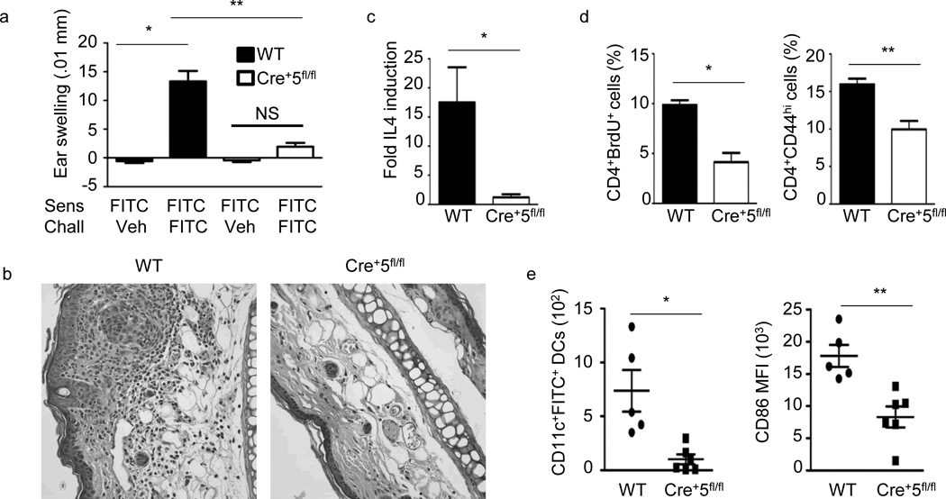
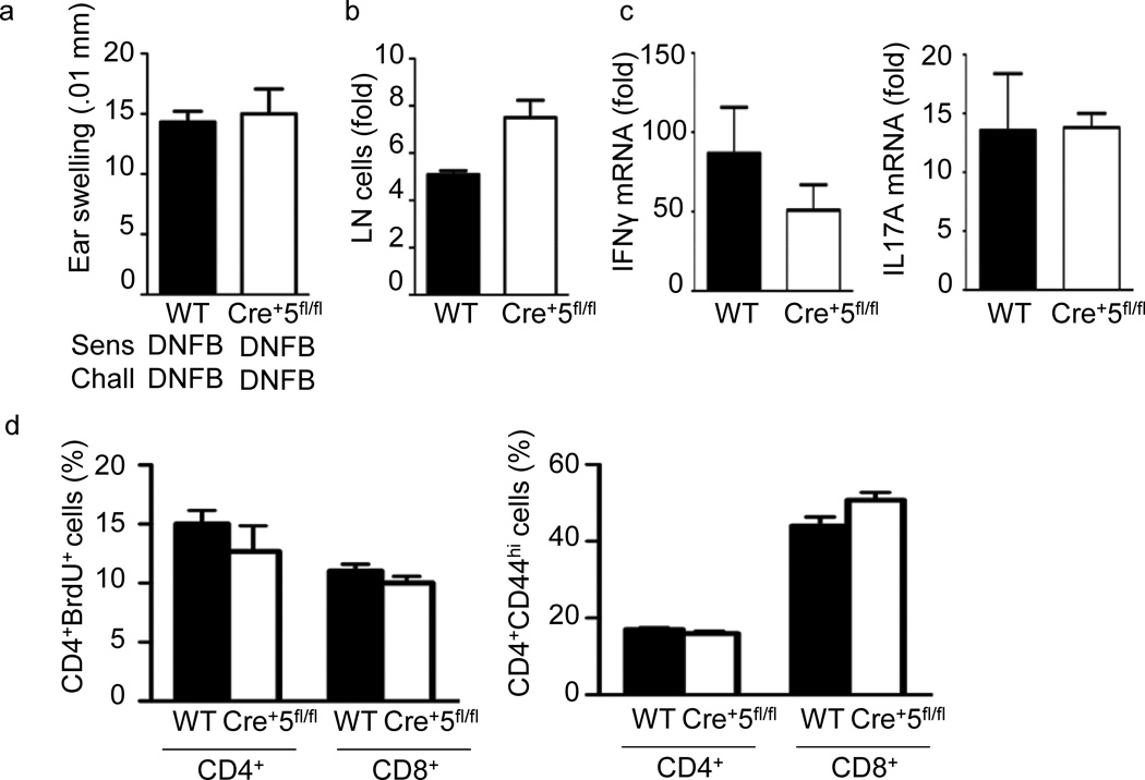
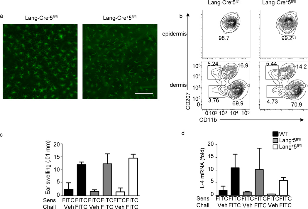
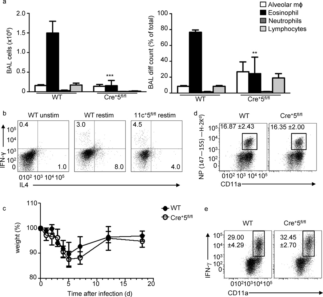
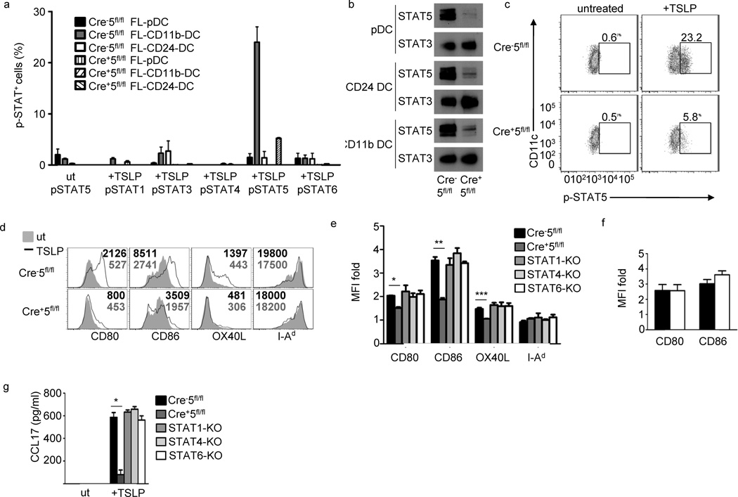
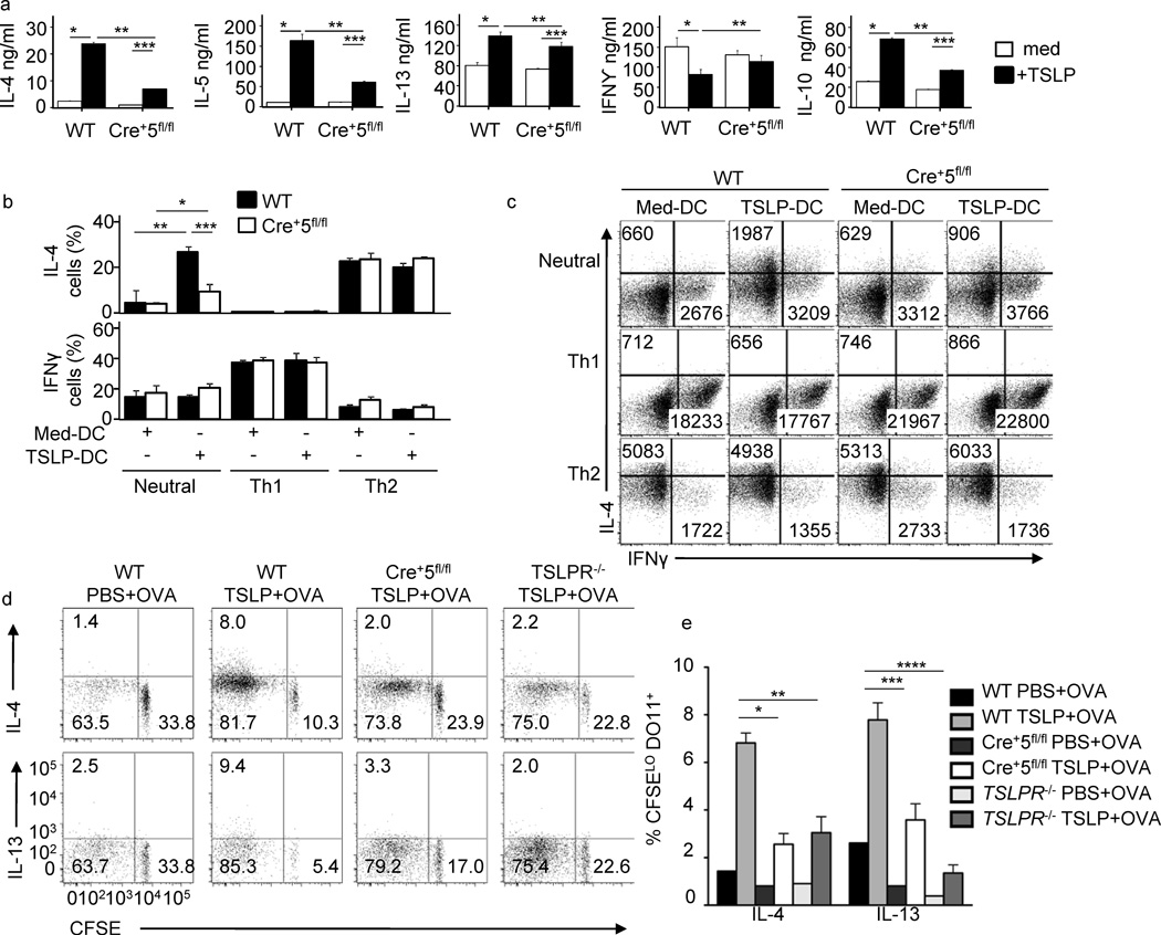
Dendritic cells (DCs) are critical in immune responses, linking innate and adaptive immunity. We found here that DC-specific deletion of the transcription factor STAT5 was not critical for development but was required for T helper type 2 (TH2), but not TH1, allergic responses in both the skin and lungs. Loss of STAT5 in DCs led to the inability to respond to thymic stromal lymphopoietin (TSLP). STAT5 was required for TSLP-dependent DC activation, including upregulation of the expression of costimulatory molecules and chemokine production. Furthermore, TH2 responses in mice with DC-specific loss of STAT5 resembled those seen in mice deficient in the receptor for TSLP. Our results show that the TSLP-STAT5 axis in DCs is a critical component for the promotion of type 2 immunity at barrier surfaces.


Figures

Figure 1
Figure 1
TH2-type CHS is significantly reduced in Cre+5fl/fl mice. (a) Mice were sensitized on the abdomen with 0.5% FITC on day 0, baseline ear thickness was measured on day 6, followed by challenge with FITC or vehicle. Change in ear thickness was measured 24 h post-challenge. WT is BALB/c control mice. * = .0001, ** = .0003. (b)Representative of five H&E-stained ear sections fixed 24 h post-challenge from WT and Cre+5fl/fl mice sensitized and challenged with FITC. (c) Ear tissue was excised and processed 24 h after challenge. IL-4 mRNA fold induction is relative to the WT vehicle-challenged samples and normalized to HPRT. * =.024 (d) Percent of CD4+ T cells positive for BrdU or CD44 from skin draining LNs 24 h following challenge. * =.005, ** = .012 (e) Number of CD11c+FITC+ DCs and the MFI of CD86 on the CD11c+FITC+ DCs in axillary and inguinal LNs 24 h post-sensitization on the abdomen with 0.5% FITC. Representative of 5 independent experiments with 4–6 mice per group, +/−SEM. * = .049, ** = .005.
Figure 2
Figure 2
Normal TH1-type CHS in Cre+5fl/fl mice. (a) Mice were sensitized with DNFB on the abdomen on day 0, baseline ear thickness was measured on day 6, followed by challenge with DNFB on the ear. Change in ear thickness was measured 24 h post-challenge. (b)Fold change in pooled axillary and inguinal LN cellularity from WT(BALB/c) and Cre+5fl/fl mice following challenge compared to non-sensitized, non-challenged mice. (c) Ear tissue was excised and processed 24 h after challenge. mRNA fold induction is relative to the WT vehicle-challenged samples and normalized to HPRT. (d)Percent of CD4+ T cells positive for BrdU or CD44 from skin dLNs 24 h following challenge. CHS results are representative of three independent experiments with 4–5 mice per group, +/-SEM.
Figure 3
Figure 3
STAT5 is not required in Langerhans cells for TH2-type CHS. (a)Fluorescent microscopy for MHCII in epidermal sheets from Lang-Cre and Lang-Cre+ STAT5fl/fl ears. Scale bar is 100µm, representative of 5 independent mice. (b)MHCII+ cells were analyzed for the expression of CD207 (langerin) and CD11b in the epidermis and dermis of Cre5fl/fl and Cre+5fl/fl mice. Graphs and percentages are representative of three independent experiments. (c) Mice were sensitized on the abdomen with 0.5% FITC on day 0, baseline ear thickness was measured on day 6, followed by challenge with FITC or vehicle. Change in ear thickness was measured 24 hours post-challenge. WT is BALB/c control mice. (d) Ear tissue was excised and processed 24 hours after challenge. IL-4 mRNA fold induction is relative to the WT vehicle-challenged samples and normalized to HPRT. Representative of three independent experiments with 3–4 mice per group.
Figure 4
Figure 4
TH2-, but not TH1-type immune responses in the lung require STAT5 in DCs. (a) Mice were sensitized with OVA-alum on day 0, followed by intranasal challenge with OVA on days 7–9, and analyzed on day 10.BAL cell number and percent of each cell population ()are average of three mice per experiment. * = .0001 (b) mLNT cells were restimulated for 4 h, followed by intracellular cytokine staining for IL-4, IL-13 and IFN-γ. ** = .0005. Wild-type and Cre+5fl/fl mice were intranasally infected with Influenza A/Aichi/68 (H3N2; 5×10³ TCID50) and (c) their weight loss was tracked as a read out of pathology. On day 8 post-infection a cohort of wild-type and Cre+5fl/fl mice were analyzed for their (d) antigen-specific CD8+ T cell response using the NP147–155/H-2Kd tetramer and (e) the percentage of influenza stimulated CD4+ T cells capable of producing IFN-γ after in vitro restimulation with PMA and ionomycin (see Methods).
Figure 5
Figure 5
Representative of 2 experiments with 5 mice per group. Figure 5 STAT5 is required for TSLP-induced costimulatory molecule upregulation in FL-CD11b-DCs. (a) Phospho-Flow of indicated STAT proteins in three FL-DC populations, in triplicate, following 15 min incubation with 50ng/ml TSLP from WT (Cre5fl/fl) and Cre+5fl/fl BMDCs. Three independent experiments. (b) Immunoblot of total-STAT5 and –STAT3 in FL-DC populations. (c) pSTAT5 phospho-flow of WT and Cre+5fl/fl FL-CD11b-DC following 15 min incubation with TSLP. (d) Costimulatory molecule expression in TSLP-treated and –untreated FL-CD11b-DCs. Numbers represent MFI values. (e) Fold-change of TSLP-treated versus untreated FL-CD11b-DCs from WT, Cre+5fl/fl, STAT-1, -4, and -6 KO bone marrow in triplicate, 4 independent experiments. * = .01, ** = .0005, *** = .05(f) Fold change of costimulatory molecule upregulation in WT (black bar) and Cre+5fl/fl (white bar) following LPS stimulation in triplicate. (g) Expression of CCL17 in FL-CD11b-DCs from e. * = .00003.
Figure 6
Figure 6
TSLP-induced STAT5 in DCs is required for TH2 differentiation of CD4+ T cells. (a) DO11.10 CD4+ T cells were cocultured with OVA(323–339)-pulsed (0.01 µM) TSLP-pretreated or untreated Cre5fl/fl and Cre+5fl/fl FL-CD11b-DCs. DCs were treated overnight with TSLP, followed by extensive washing to remove residual TSLP before addition of T cells. T cells were stimulated for 5 days, followed by ELISA for indicated cytokines. *P < 0.05 and **P < 0.01 (one-way ANOVA). (b, c) Percent CD4+ T cells positive for IL-4 and IFN-γ (b, bar graphs) and average MFI for×and y axis separately. *P < 0.05 and **P < 0.01 (one-way ANOVA). c, numbers in FACs plotsafter coculture with TSLP-pretreated and untreated Cre5fl/fl and Cre+5fl/fl FL-CD11b-DCs in neutral, TH1 and TH2 skewing conditions, measured by flow cytometry. (d) WT, Cre+5fl/fl and TSLPR−/− were intravenously injected with CFSE labeled DO11.10 naïve CD4+ T cells. Mice were given intradermal injections of OVA plus or minus TSLP in PBS. Seven days later, pooled skin dLNs were restimulated and intracellular cytokine staining for IL-13 and IL-4 was detected by flow cytometry and graphed relative to CFSE dilution. Representative of four mice per group, two independent experiments. (e) Percent of CFSEloDO11.10+ T cells from (d) positive for IL-4 or IL-13. * = .002, ** = .003, *** = .001, **** = .0003.

References

    1. Shortman K, Naik SH. Steady-state and inflammatory dendritic-cell development. Nat Rev Immunol. 2007;7:19–30. - PubMed
    1. McKenna HJ, et al. Mice lacking flt3 ligand have deficient hematopoiesis affecting hematopoietic progenitor cells, dendritic cells, and natural killer cells. Blood. 2000;95:3489–3497. - PubMed
    1. Laouar Y, Welte T, Fu XY, Flavell RA. STAT3 is required for Flt3L-dependent dendritic cell differentiation. Immunity. 2003;19:903–912. - PubMed
    1. Onai N, Obata-Onai A, Tussiwand R, Lanzavecchia A, Manz MG. Activation of the Flt3 signal transduction cascade rescues and enhances type I interferon-producing and dendritic cell development. J Exp Med. 2006;203:227–238. - PMC - PubMed
    1. Esashi E, et al. The signal transducer STAT5 inhibits plasmacytoid dendritic cell development by suppressing transcription factor IRF8. Immunity. 2008;28:509–520. - PMC - PubMed

Publication types

MeSH terms