. 2013 Apr;12(4):627-34.
doi: 10.1128/EC.00016-13. Epub 2013 Feb 22.

Structural insights into substrate binding by PvFKBP35, a peptidylprolyl cis-trans isomerase from the human malarial parasite Plasmodium vivax

Affiliations

Structural insights into substrate binding by PvFKBP35, a peptidylprolyl cis-trans isomerase from the human malarial parasite Plasmodium vivax

Reema Alag et al. Eukaryot Cell. 2013 Apr.

Abstract

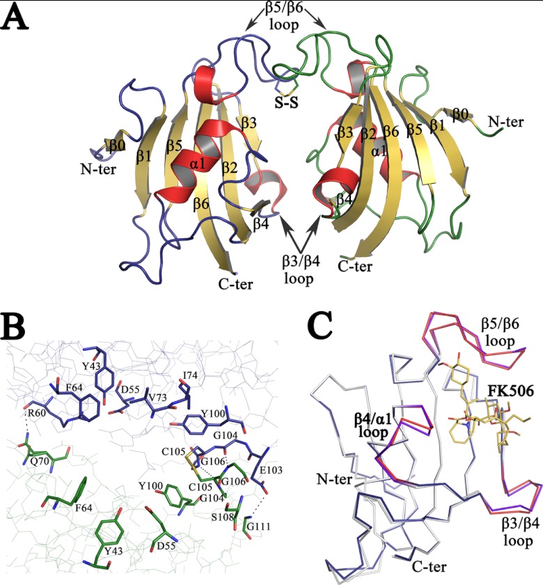
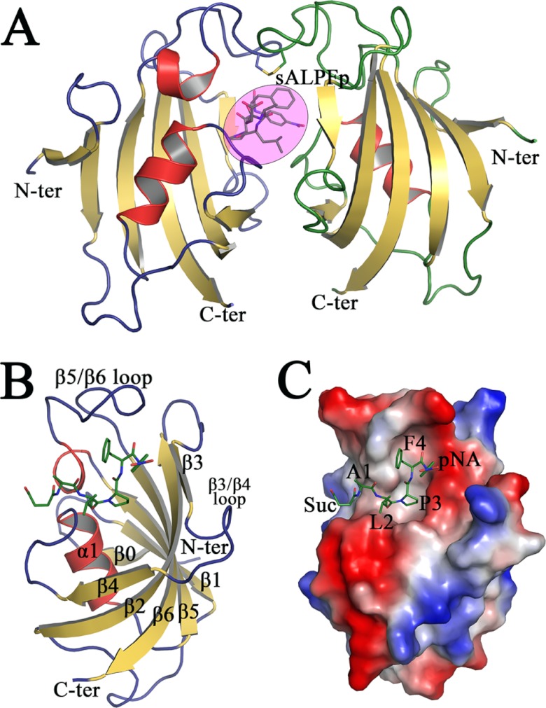
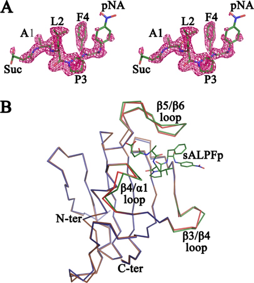
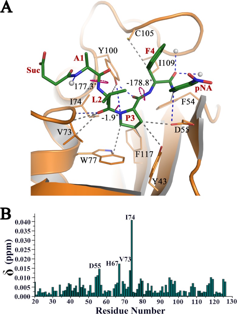
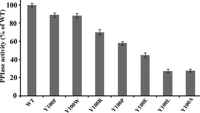
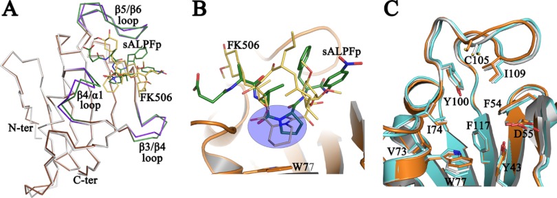
The immunosuppressive drug FK506 binding proteins (FKBPs), an immunophilin family with the immunosuppressive drug FK506 binding property, exhibit peptidylprolyl cis-trans isomerase (PPIase) activity. While the cyclophilin-catalyzed peptidylprolyl isomerization of X-Pro peptide bonds has been extensively studied, the mechanism of the FKBP-mediated peptidylprolyl isomerization remains uncharacterized. Thus, to investigate the binding of FKBP with its substrate and the underlying catalytic mechanism of the FKBP-mediated proline isomerization, here we employed the FK506 binding domain (FKBD) of the human malarial parasite Plasmodium vivax FK506 binding protein 35 (PvFKBP35) and examined the details of the molecular interaction between the isomerase and a peptide substrate. The crystallographic structures of apo PvFKBD35 and its complex with the tetrapeptide substrate succinyl-Ala-Leu-Pro-Phe-p-nitroanilide (sALPFp) determined at 1.4 Å and 1.65 Å resolutions, respectively, showed that the substrate binds to PvFKBD35 in a cis conformation. Nuclear magnetic resonance (NMR) studies demonstrated the chemical shift perturbations of D55, H67, V73, and I74 residues upon the substrate binding. In addition, the X-ray crystal structure, along with the mutational studies, shows that Y100 is a key residue for the catalytic activity. Taken together, our results provide insights into the catalytic mechanism of PvFKBP35-mediated cis-trans isomerization of substrate and ultimately might aid designing substrate mimetic inhibitors targeting the malarial parasite FKBPs.


Figures

Fig 1
Fig 1
Crystal structure of apo PvFKBD35. (A) The overall structure of the FKBD35 domain of P. vivax is shown as a cartoon diagram. The disulfide bond between Cys 105 of both chains is displayed in stick mode with the sulfur atoms colored in yellow. The loops corresponding to chains A (left) and B (right) are colored blue and green, respectively, while the helices are colored red and sheets are in yellow. (B) Close view of active-site residues, in stick mode, that line up the dimer interface formed between the two PvFKBD35 (chain A, blue; and chain B, green) molecules in the asymmetric unit. The disulfide bond is shown as a stick, and hydrogen bonds are displayed as dashed lines. (C) Shown is the superimposition of the backbone traces of apo PvFKBD35 colored in blue (β3-β4, β4-α1, and β5-β6 loops colored red) and FK506-bound PvFKBD35 colored in white (β3-β4, β4-α1, and β5-β6 loops colored purple). The FK506 inhibitor is shown as yellow-colored sticks.
Fig 2
Fig 2
Crystal structure of sALPFp-bound PvFKBD35. (A) The cartoon diagram of the two monomers of PvFKBD35-sALPFp in the asymmetric unit, with color code and orientation similar to those described in Fig. 1A. The sALPFp tetrapeptide is represented as sticks within the pink-colored circle. (B) Cartoon representation of the complex formed between PvFKBD35 and sALPFp (in green sticks), with the secondary structures labeled in black. (C) Electrostatic surface potential representation of PvFKBD35, with sALPFp shown as green-colored sticks. It could be seen that the proline of sALPFp docks itself deep into the hydrophobic pocket.
Fig 3
Fig 3
Stereoview of sALPFp peptide and overlay of apo PvFKBD35 and sALPFp-bound PvFKBD35. (A) Stereoview of the electron density map (2FO-FC) contoured at the 1.0 σ level for the peptide molecule. It could be clearly seen that the Leu-Pro peptide bond adopts a cis-isomer conformation. (B) Superposition of the Cα traces of apo PvFKBD35 colored in blue and PvFKBD35 bound to sALPFp colored in brown. The ligand-flanking loops of the apo- and sALPFp-bound structures are colored in red and green, respectively. The sALPFp peptide is shown as green sticks for reference.
Fig 4
Fig 4
Interaction of sALPFp with PvFKBD35. (A) The hydrogen bonded (blue dashes) and nonbonded (gray dashes) interactions made by sALPFp (green sticks) with PvFKBD35, revealing the binding of the proline (P3) region into the deep active-site pocket. The three ω angles corresponding to the peptide bonds are also labeled and shown by pink dots. (B) Chemical shift perturbations of PvFKBD35 (0.35 mM) upon adding the substrate peptide. Residues showing perturbations upon addition of the peptide are labeled.
Fig 5
Fig 5
PPIase activity of wild-type PvFKBD35 and the Y100 mutants. PPIase assay was performed at 4°C as described in Materials and Methods, and change in the absorbance was measured at 390 nm for 5 min.
Fig 6
Fig 6
Comparison of FK506 and sALPFp binding to PvFKBD35. (A) Shown is a superposition of the Cα traces of FK506-bound PvFKBD35 (colored in white) with the sALPFp complex (colored in brown). The ligand-flanking loops of the FK506- and sALPFp-bound structures are colored in purple and green, respectively. The bound FK506 and sALPFp are represented in yellow and green sticks, respectively. (B) A closer view of the superposition of the FK506 (yellow sticks) over the sALPFp (green sticks) shows that the pipecolic moiety of FK506 overlays itself on the proline residue of substrate (within the blue circle). The cartoon representation of the PvFKBD35-sALPFp (in orange) and W77 (in stick mode), which forms the base of the pocket, is shown for reference. (C) The common residues that are involved in the binding of FK506 (in cyan) and sALPFp (in orange) in the PvFKBD35 structure are shown in stick mode, while those of the apo structure are shown in white. The figure enables us to visualize the subtle changes in the active-site residues due to ligand binding.

References

    1. Fischer G, Aumuller T. 2003. Regulation of peptide bond cis/trans isomerization by enzyme catalysis and its implication in physiological processes. Rev. Physiol. Biochem. Pharmacol. 148:105–150 - PubMed
    1. Lu KP, Hanes SD, Hunter T. 1996. A human peptidyl-prolyl isomerase essential for regulation of mitosis. Nature 380:544–547 - PubMed
    1. Ou W, Luo W, Park YD, Zhou HM. 2001. Chaperone-like activity of peptidyl-prolyl cis-trans isomerase during creatine kinase refolding. Protein Sci. 10:2346–2353 - PMC - PubMed
    1. Lu KP, Finn G, Lee TH, Nicholson LK. 2007. Prolyl cis-trans isomerization as a molecular timer. Nat. Chem. Biol. 3:619–629 - PubMed
    1. Fischer G. 2000. Chemical aspects of peptide bond isomerization. Chem. Soc. Rev. 29:119–127

Publication types

MeSH terms

LinkOut - more resources