. 2013 May;79(9):2968-78.
doi: 10.1128/AEM.03666-12. Epub 2013 Feb 22.

Role of PelF in pel polysaccharide biosynthesis in Pseudomonas aeruginosa

Affiliations

Role of PelF in pel polysaccharide biosynthesis in Pseudomonas aeruginosa

Aamir Ghafoor et al. Appl Environ Microbiol. 2013 May.

Abstract

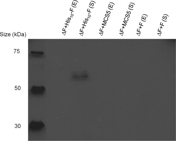
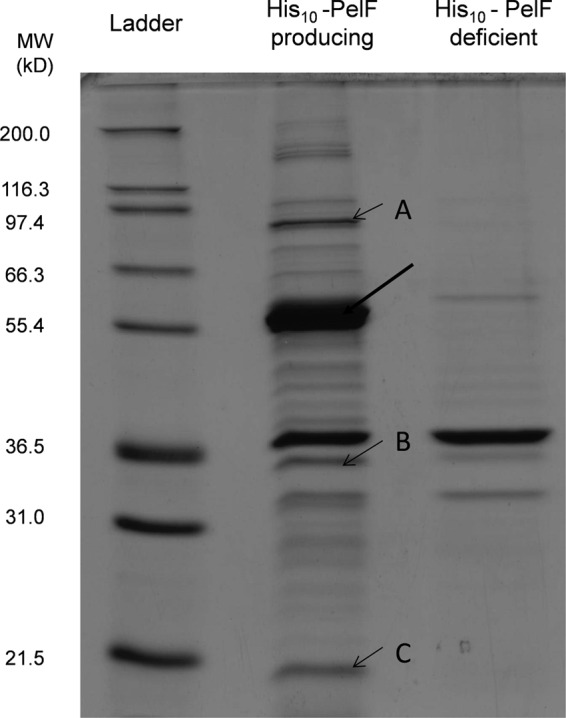
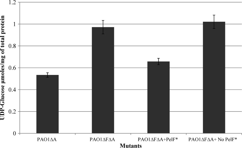
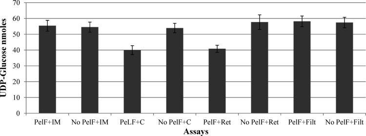
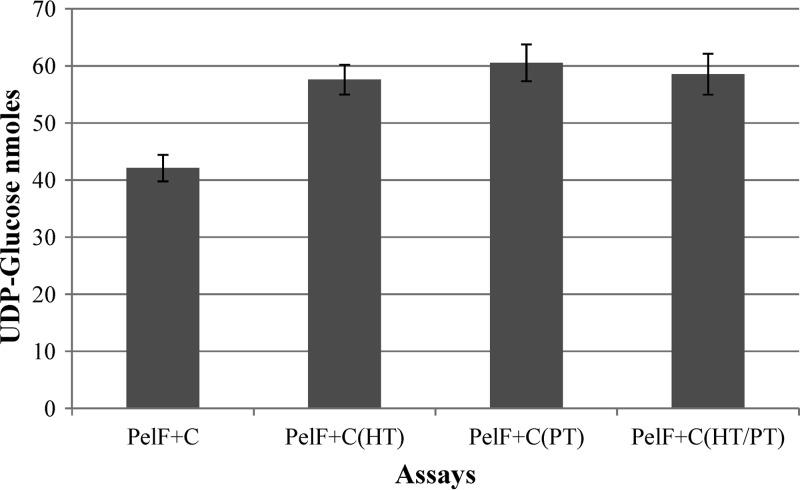
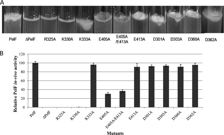
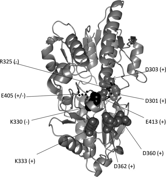
Pseudomonas aeruginosa produces three exopolysaccharides, Psl, Pel, and alginate, that play vital roles in biofilm formation. Pel is a glucose-rich, cellulose-like exopolysaccharide. The essential Pel biosynthesis proteins are encoded by seven genes, pelA to pelG. Bioinformatics analysis suggests that PelF is a cytosolic glycosyltransferase. Here, experimental evidence was provided to support this PelF function. A UDP-glucose dehydrogenase-based assay was developed to quantify UDP-glucose. UDP-glucose was proposed as the substrate for PelF. The isogenic pelF deletion mutant accumulated 1.8 times more UDP-glucose in its cytosol than the wild type. This suggested that PelF, which was found localized in the cystosol, uses UDP-glucose as substrate. Additionally, in vitro experiments confirmed that PelF uses UDP-glucose as substrate. To analyze the functional roles of conserved residues in PelF, site-directed mutagenesis was performed. The presence of the EX7E motif is characteristic for various glycosyltransferase families, and in PelF, E405/E413 are the conserved residues in this motif. Replacement of E405 with A resulted in a reduction of PelF activity to 30.35% ± 3.15% (mean ± standard deviation) of the wild-type level, whereas replacement of the second E, E413, with A did not produce a significant change in the activity of PelF. Moreover, replacement of both E residues did not result in a loss of PelF function, but replacement of the conserved R325 or K330 with A resulted in a complete loss of PelF activity. Overall, our data show that PelF is a soluble glycosyltransferase that uses UDP-glucose as the substrate for Pel synthesis and that conserved residues R325 and K330 are important for the activity of PelF.


Figures

Fig 1
Fig 1
Immunoblot results using anti-His antibodies to determine the subcellular localization of PelF. Soluble (S) and envelope (E) fractions of P. aeruginosa PAO1 ΔpslA ΔpelF harboring pBBR1-MCS-5::His10-PelF, empty vector (pBBR1-MCS-5), and pBBR1-MCS-5::PelF (without His tag) were subjected to SDS-PAGE.
Fig 2
Fig 2
SDS-PAGE analysis of partially purified PelF. Soluble fractions of PelF-producing and PelF-deficient strains were subjected to a HSMP column, and elution fractions were analyzed by SDS-PAGE. An arrow (thick) indicates the presence of His10-PelF. Three proteins coeluting with PelF were identified: A, ATP-dependent protease (GI 15595976); B, PA4657 (GI 15599852) of P. aeruginosa; C, Gcn5-related N-acetyltransferase (GI 239721) (best match to Serratia marcescens).
Fig 3
Fig 3
Concentration of UDP-glucose in the soluble fraction of various P. aeruginosa PAO1 mutants. PAO1ΔA (PAO1 ΔpslA), Psl-deficient/Pel-producing mutant; PAO1ΔFΔA (PAO1 ΔpelFΔpslA), Pel-deficient/Psl-deficent mutant; PAO1ΔFΔA (PAO1 ΔpelFΔpslA) + PelF, partially purified PelF added to soluble fraction of cell lysate from Pel-deficient/Psl-deficent mutant (*PelF was partially purified as described in Materials and Methods); PAO1ΔFΔA (PAO1 ΔpelFΔpslA) + No PelF, the elution fraction not containing PelF but containing coeluted proteins was added to the soluble fraction of the cell lysate from the Pel-deficient/Psl-deficient mutant. All experiments were conducted in triplicate, and mean values presented in the graphs. Standard deviations are shown as the error bars.
Fig 4
Fig 4
PelF activity as measured by UDP-glucose consumption. The residual UDP-glucose after reaction completion was determined. PelF+IM, PelF and 5 μl IM fraction (containing 4 mg/ml total protein); no PeLF+IM, partially purified suspension without PelF and 5 μl (containing 4 mg/ml total protein) inner membrane fraction; PelF+C, PelF and 5 μl soluble fraction (containing 4 mg/ml total protein); no PeLF+C, partially purified suspension without PelF and 5 μl soluble fraction (containing 4 mg/ml total protein); PelF+Ret, PelF and 5 μl retentate of soluble fraction (containing 4 mg/ml total protein) filtered; no PeLF+Ret, partially purified suspension without PelF and 5 μl retentate of soluble fraction (containing 4 mg/ml total protein); PelF+Filt, PelF and 5 μl filtrate of soluble fraction (containing 1 mg/ml total protein); no PeLF+Filt, partially purified suspension without PelF and 5 μl filtrate of soluble fraction (containing 1 mg/ml total protein). All experiments were conducted in triplicate, and mean values are presented. Standard deviations are shown as error bars.
Fig 5
Fig 5
PelF activity measured by UDP-glucose consumption. The residual UDP-glucose was measured after completion of the assay. PelF+C, PelF and 5 μl untreated soluble fraction (containing 4 mg/ml total protein); PelF+C (HT), PelF and 5 μl heat-treated soluble fraction; PelF+C(PT), PelF and 5 μl protease-treated soluble fraction; PelF+C (HT/PT), PelF and 5 μl protease- and heat-treated soluble fraction.
Fig 6
Fig 6
In vivo activities of PelF and its variants as determined by pellicle production at the air-liquid interface by various P. aeruginosa strains. Pellicle produced by P. aeruginosa strains harboring different variants of PelF after 96-h static cultures was quantified using the Congo red binding assay as described in Materials and Methods. (A) Congo red binding assay results. The percentages shown are the mean values of three independent assays. Standard deviations are presented as error bars. (B) Pellicle formation mediated by variants of PelF, compared with that of wild-type His10-PelF.
Fig 7
Fig 7
Structural model of PelF generated by PHYRE2, showing localization of the amino acid residues that were replaced with alanine. Selected amino acid residues were replaced with alanine by using site-directed mutagenesis as described in Materials and Methods. Residues targeted for replacement are shown as spheres. +, no impact on activity; +/−, partial loss of enzymatic activity; −, activity of the enzyme was lost.

References

    1. Singh PK, Schaefer AL, Parsek MR, Moninger TO, Welsh MJ, Greenberg EP. 2000. Quorum-sensing signals indicate that cystic fibrosis lungs are infected with bacterial biofilms. Nature 407: 762–764 - PubMed
    1. Drenkard E. 2003. Antimicrobial resistance of Pseudomonas aeruginosa biofilms. Microbes Infect. 5: 1213–1219 - PubMed
    1. Ghafoor A, Hay ID, Rehm BH. 2011. Role of exopolysaccharides in Pseudomonas aeruginosa biofilm formation and architecture. Appl. Environ. Microbiol. 77: 5238–5246 - PMC - PubMed
    1. Chitnis CE, Ohman DE. 1993. Genetic analysis of the alginate biosynthetic gene cluster of Pseudomonas aeruginosa shows evidence of an operonic structure. Mol. Microbiol. 8: 583–593 - PubMed
    1. Friedman L, Kolter R. 2004. Genes involved in matrix formation in Pseudomonas aeruginosa PA14 biofilms. Mol. Microbiol. 51: 675–690 - PubMed

Publication types

MeSH terms

LinkOut - more resources